Physiology notes
Excitation of the skeletal muscles
In our spine (CNS): somatic alpha motor neurons that secrete ACh. Release
of ACh always leads to contraction, and no antagonistic innervation.
Always excitatory!
ACh depolarizes the muscle membrane (nicotinic receptor) muscle
contraction.
Depending on the body part: number of neurons innervating.
A motor unit: a somatic motor neuron plus all the muscle fibres it
stimulates.
*note: medulla of the adrenal is a modified ganglion.
Neuromuscular junction
Axons are branching close to the muscle fibres, creating the
neuromuscular junction. One somatic motor neuron can innervate
multiple muscle fibres. One muscle fibre cannot get information from
multiple neurons.
Neuromuscular junction: axon terminals, motor end plates on the muscle
membrane and Schwann cell sheaths. A motor end plate is a region of
muscle membrane (specialisation of the muscle membrane) that contains
high concentrations of nicotinic ACh receptors (ionotropic).
- A muscle fibre is a single cell.
- A muscle fibre has multiple nuclei, caused by the fusion of muscle
cells.
- Schwann cells: velocity action potential, development junction,
growth factors.
- The synaptic cleft is collagen filled to make the connection very
tight.
- Depolarization of the membrane opens voltage gated Ca2+
channels. Ca2+ enters the cell to open synaptic vesicles with ACh.
- The enzyme Acetylcholinesterase (AChE) breaks down ACh at the
end of the membrane.
When ACh binds to the receptor, gates open and Na+ travels into the cell
and K+ travels into the cell. There is an overflow of Na+ into the cell,
which causes the depolarization of the membrane contraction of muscle
fibre.
- Relaxation: inhibition at the level of somatic alpha motor neurons in
CNS. They can be stopped firing causing relaxation.
- Exercise facilitates recovery after damage to innervating axons.
Regeneration of muscle cells when damaged by satellite cells. Satellite
cells get activated when there is damage. These cells merge to form a
muscle fibre. Only when young!
When older: deficient regeneration (loss of polarity, apoptosis etc.)
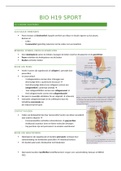
Types of muscles
- Skeletal muscles: large fibres, multinucleate cells
are striped/striated under the microscope.
, - Cardiac muscle: also striated fibres but they are smaller, branched
and uninucleate. Cells are joined in series by junction (intercalated
disks).
- Smooth muscle (blood vessels): fibres are small and lack striations.
Skeletal muscle Smooth & heart muscle
Locomotor movement Movement of content (like blood)
Connected to bone Not connected to bone
Voluntary Involuntary
Somatic motor neuron: excitation Multiple control (excitation-
inhibition: autonomic nervous,
intrinsic, endocrine)
Movement via joints:
- Flexor (moves body parts
toward)
- Extensor (moves body parts
away)
Muscle cell = muscle fibre
Cell membrane = sarcolemma
Cytoplasm = sarcoplasm
Modified ER = sarcoplasmic reticulum
Sarcomere = functional unit of striated muscle tissue.
T-tubules are extensions of the muscle membrane into the muscle fibre.
Depolarization must travel rapidly over the muscle fibre reaching all the
myofibrils. T-tubules make this possible. Myofibrils contain thick and thin
filaments (contractile proteins), thick pulls thin inside by myosin heads.
Thick filaments: myosin. Thin filaments: actin.
Sarcoplasmic reticulum contains Calcium. Outside of the SR touches the
membrane, this close contact is necessary for the release of calcium.
Regulatory proteins: tropomyosin and
troponin = allow the pulling to happen.
Attached to the actin filaments.
‘Giant accessory’ proteins: titin
stabilizes the thick filaments and holds
elasticity. Largest protein in your body.
Myosin molecules: Hinge region and 2
myosin heads (motor protein)
Excitation of the skeletal muscles
In our spine (CNS): somatic alpha motor neurons that secrete ACh. Release
of ACh always leads to contraction, and no antagonistic innervation.
Always excitatory!
ACh depolarizes the muscle membrane (nicotinic receptor) muscle
contraction.
Depending on the body part: number of neurons innervating.
A motor unit: a somatic motor neuron plus all the muscle fibres it
stimulates.
*note: medulla of the adrenal is a modified ganglion.
Neuromuscular junction
Axons are branching close to the muscle fibres, creating the
neuromuscular junction. One somatic motor neuron can innervate
multiple muscle fibres. One muscle fibre cannot get information from
multiple neurons.
Neuromuscular junction: axon terminals, motor end plates on the muscle
membrane and Schwann cell sheaths. A motor end plate is a region of
muscle membrane (specialisation of the muscle membrane) that contains
high concentrations of nicotinic ACh receptors (ionotropic).
- A muscle fibre is a single cell.
- A muscle fibre has multiple nuclei, caused by the fusion of muscle
cells.
- Schwann cells: velocity action potential, development junction,
growth factors.
- The synaptic cleft is collagen filled to make the connection very
tight.
- Depolarization of the membrane opens voltage gated Ca2+
channels. Ca2+ enters the cell to open synaptic vesicles with ACh.
- The enzyme Acetylcholinesterase (AChE) breaks down ACh at the
end of the membrane.
When ACh binds to the receptor, gates open and Na+ travels into the cell
and K+ travels into the cell. There is an overflow of Na+ into the cell,
which causes the depolarization of the membrane contraction of muscle
fibre.
- Relaxation: inhibition at the level of somatic alpha motor neurons in
CNS. They can be stopped firing causing relaxation.
- Exercise facilitates recovery after damage to innervating axons.
Regeneration of muscle cells when damaged by satellite cells. Satellite
cells get activated when there is damage. These cells merge to form a
muscle fibre. Only when young!
When older: deficient regeneration (loss of polarity, apoptosis etc.)
Types of muscles
- Skeletal muscles: large fibres, multinucleate cells
are striped/striated under the microscope.
, - Cardiac muscle: also striated fibres but they are smaller, branched
and uninucleate. Cells are joined in series by junction (intercalated
disks).
- Smooth muscle (blood vessels): fibres are small and lack striations.
Skeletal muscle Smooth & heart muscle
Locomotor movement Movement of content (like blood)
Connected to bone Not connected to bone
Voluntary Involuntary
Somatic motor neuron: excitation Multiple control (excitation-
inhibition: autonomic nervous,
intrinsic, endocrine)
Movement via joints:
- Flexor (moves body parts
toward)
- Extensor (moves body parts
away)
Muscle cell = muscle fibre
Cell membrane = sarcolemma
Cytoplasm = sarcoplasm
Modified ER = sarcoplasmic reticulum
Sarcomere = functional unit of striated muscle tissue.
T-tubules are extensions of the muscle membrane into the muscle fibre.
Depolarization must travel rapidly over the muscle fibre reaching all the
myofibrils. T-tubules make this possible. Myofibrils contain thick and thin
filaments (contractile proteins), thick pulls thin inside by myosin heads.
Thick filaments: myosin. Thin filaments: actin.
Sarcoplasmic reticulum contains Calcium. Outside of the SR touches the
membrane, this close contact is necessary for the release of calcium.
Regulatory proteins: tropomyosin and
troponin = allow the pulling to happen.
Attached to the actin filaments.
‘Giant accessory’ proteins: titin
stabilizes the thick filaments and holds
elasticity. Largest protein in your body.
Myosin molecules: Hinge region and 2
myosin heads (motor protein)