NEUROLOGICAL ASPECTS:
NEURAL NETWORKS AND
REORGANIZATION
Academiejaar 2023-2024 – 2MARE
H1. NEUROSCIENTIFIC METHODS
H2. THE POTENTIAL OF NON-INVASIVE BRAIN
STIMULATION IN NEUROREHABILITATION
H3. AGEING AND NEUROPLASTICITY
H4. NEUROREHABILITATION AFTER STROKE
PART PROF. KAAT ALAERTS
, PART KAAT ALAERTS
Neurological aspects: neural networks
and reorganization
CHAPTER 1: NEUROSCIENTIFIC METHODS
• Measuring ‘brain activity’ at the systems level (not single-cell)
• Non-invasive (not ‘inside’ the brain, but at the level of the scull)
• Fundamental research
o Motor control, motor learning
o Cognition
o Memory
o ...
• Clinical research
o Neural processes underlying ageing
o Neural basis of diseases (stroke, Parkinson, epilepsy, neurodevelopmental disorders)
o Neural evaluation of disease progression
o Neural evaluation of interventions/ treatments
Look for each technique at equipment, neurophysiological basis and examples of applications
Important concept to evaluate the advantages and disadvantages of different techniques
Temporal and spatial resolution
o Some better for temporal: what is happening in time regarding brain activity?
o TEMPORAL = when in time
o SPATIAL = where in the brain
1/ MAGNETIC RESONANCE IMAGING (MRI)
What isn’t fMRI?
➢ fMRI is not bumpology (claims that bumps on the skull reflected exaggerated functions/traits)
➢ fMRI is not mind-reading
➢ fMRI is not invasive (the skull remains closed)
What is fMRI: a relative, safe, non-invasive technique
1
, PART KAAT ALAERTS
1.1 Equipment
- Giant magnet
- Head coil (helps with high-quality images)
MRI = brain anatomy
fMRI = functional MRI = brain activity
fcMRI = functional connectivity
First MRI-scan around 1950, first functional MRI-scan developed way later (around 1990)
1.2 Biological basis of MRI
• Measures brain anatomy
• Former name: (Nuclear) Magnetic Resonance Imaging → nothing to do with ‘radioactivity’, but
with the magnetic properties of protons, in the nuclei of atoms
Protons:
- Have a mass, are positive (+) and have a spin (they turn around)
- Because protons turn around, they have a small, but measurable magnetic field
- In everyday life, the protons in our body are in balance, randomly oriented, but in balance
- Inside the MRI scanner, which is one giant magnet, the protons align to the magnetic field (B0).
Either in parallel (same direction) or anti-parallel (opposite direction)
- Most protons will align parallel in MRI-scan
- The majority of atoms aligns in parallel, allowing to define the NET magnetization of protons
in the direction of B0
- This is what happens if you’re positioned in the scanner. And this magnetic field is ALWAYS on.
- Emission of a radio frequency pulse by the head coil, induces a flip of the NET magnetization
(instead of aligning to the Z- axis, the protons now align in the X-Y field)
- Proton is in excitation state
- Head coils will emit a radiofrequency: magnetic field of protons will shift (excitation
state) → follow the Y-axis
- However, protons don’t like being in this ‘high-energy’ excitation state’, and from the moment
the radio frequency pulse is turned off, it will ‘relax’ to its initial position (i.e., align back to the
Z-axis of the B0 field).
- During this ‘relaxation state’, the protons emit radio frequency themselves, and this signal is
measured. (The head coil, both emits and measures radio frequencies)
- Proton emits radio frequency during relaxation state
- Protons want to align with the Z-axis → when the radiofrequency is turned off, they
return to the Z-axis
- While doing this, they will emit radiofrequency itself → MRI can measure the different
relaxation times in the different tissue
2
, PART KAAT ALAERTS
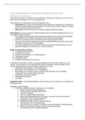
The time it takes for a proton to relax to 63% of its initial state (along the z-axis) is called T1
T1 = relaxation time
• Not all tissues ‘relax’ the same way!
• Protons in fat (e.g., white matter), relax much faster, than protons
in liquid (e.g., cerebrospinal fluid)
• By measuring the relaxation in different tissues, contrasts can be
visualized!
• In so-called ‘T1-weighted’ images, liquid is dark (less energy emitted), and fat is bright (more
energy emitted) → dark matter needs less energy at T1 compared to white matter (grey matter
is in between)
1.2.1 Examples of application
➢ Clinical: Localization of brain lesions
➢ Used in pre-surgical mapping (e.g., epilepsy) and prediction of disease progression
1.3 Functional MRI (fMRI)
Uses MRI to indirectly measure brain activity
Same principle as MRI: contrasts in brain images based on measurement of magnetic relaxation
→ Here however, it is not about protons, but about hemoglobin in the blood...
1.3.1 Biological basis of fMRI
• Brain region active => increased O2 metabolism => increased blood flow
• fMRI measures the Blood Oxygen Level Dependent (BOLD) signal
o Oxyhemoglobin = diamagnetic (same as tissue)
o Deoxyhemoglobin = paramagnetic (weak magnetic) → interacts with the signal of MRI
• fMRI always measures a change in BOLD-response
o You always need a baseline to see how BOLD has changed
o During baseline situation, the BOLD-signal goes back down again
LEFT-LEFT = brain at rest
Basic metabolism (good balance between oxy and de-oxy Hb in bloodstream, normal usage of
oxygen by the neuronal cells, normal glucoses dosage as energy resource)
LEFT-RIGHT = activation of the neuron
→ Neurons will require an increased metabolism of glucoses and oxygen
Initially in bloodstream will be higher amount of de-oxy compared to oxy
→ This gives a reduced BOLD-signal
RIGHT = this triggers a haemodynamic respons rendering an increased transport of oxy to this region
of activation
Rendering in a lowering of the ratio de-oxy/oxy → rendering in an increase of BOLD-signals
3
NEURAL NETWORKS AND
REORGANIZATION
Academiejaar 2023-2024 – 2MARE
H1. NEUROSCIENTIFIC METHODS
H2. THE POTENTIAL OF NON-INVASIVE BRAIN
STIMULATION IN NEUROREHABILITATION
H3. AGEING AND NEUROPLASTICITY
H4. NEUROREHABILITATION AFTER STROKE
PART PROF. KAAT ALAERTS
, PART KAAT ALAERTS
Neurological aspects: neural networks
and reorganization
CHAPTER 1: NEUROSCIENTIFIC METHODS
• Measuring ‘brain activity’ at the systems level (not single-cell)
• Non-invasive (not ‘inside’ the brain, but at the level of the scull)
• Fundamental research
o Motor control, motor learning
o Cognition
o Memory
o ...
• Clinical research
o Neural processes underlying ageing
o Neural basis of diseases (stroke, Parkinson, epilepsy, neurodevelopmental disorders)
o Neural evaluation of disease progression
o Neural evaluation of interventions/ treatments
Look for each technique at equipment, neurophysiological basis and examples of applications
Important concept to evaluate the advantages and disadvantages of different techniques
Temporal and spatial resolution
o Some better for temporal: what is happening in time regarding brain activity?
o TEMPORAL = when in time
o SPATIAL = where in the brain
1/ MAGNETIC RESONANCE IMAGING (MRI)
What isn’t fMRI?
➢ fMRI is not bumpology (claims that bumps on the skull reflected exaggerated functions/traits)
➢ fMRI is not mind-reading
➢ fMRI is not invasive (the skull remains closed)
What is fMRI: a relative, safe, non-invasive technique
1
, PART KAAT ALAERTS
1.1 Equipment
- Giant magnet
- Head coil (helps with high-quality images)
MRI = brain anatomy
fMRI = functional MRI = brain activity
fcMRI = functional connectivity
First MRI-scan around 1950, first functional MRI-scan developed way later (around 1990)
1.2 Biological basis of MRI
• Measures brain anatomy
• Former name: (Nuclear) Magnetic Resonance Imaging → nothing to do with ‘radioactivity’, but
with the magnetic properties of protons, in the nuclei of atoms
Protons:
- Have a mass, are positive (+) and have a spin (they turn around)
- Because protons turn around, they have a small, but measurable magnetic field
- In everyday life, the protons in our body are in balance, randomly oriented, but in balance
- Inside the MRI scanner, which is one giant magnet, the protons align to the magnetic field (B0).
Either in parallel (same direction) or anti-parallel (opposite direction)
- Most protons will align parallel in MRI-scan
- The majority of atoms aligns in parallel, allowing to define the NET magnetization of protons
in the direction of B0
- This is what happens if you’re positioned in the scanner. And this magnetic field is ALWAYS on.
- Emission of a radio frequency pulse by the head coil, induces a flip of the NET magnetization
(instead of aligning to the Z- axis, the protons now align in the X-Y field)
- Proton is in excitation state
- Head coils will emit a radiofrequency: magnetic field of protons will shift (excitation
state) → follow the Y-axis
- However, protons don’t like being in this ‘high-energy’ excitation state’, and from the moment
the radio frequency pulse is turned off, it will ‘relax’ to its initial position (i.e., align back to the
Z-axis of the B0 field).
- During this ‘relaxation state’, the protons emit radio frequency themselves, and this signal is
measured. (The head coil, both emits and measures radio frequencies)
- Proton emits radio frequency during relaxation state
- Protons want to align with the Z-axis → when the radiofrequency is turned off, they
return to the Z-axis
- While doing this, they will emit radiofrequency itself → MRI can measure the different
relaxation times in the different tissue
2
, PART KAAT ALAERTS
The time it takes for a proton to relax to 63% of its initial state (along the z-axis) is called T1
T1 = relaxation time
• Not all tissues ‘relax’ the same way!
• Protons in fat (e.g., white matter), relax much faster, than protons
in liquid (e.g., cerebrospinal fluid)
• By measuring the relaxation in different tissues, contrasts can be
visualized!
• In so-called ‘T1-weighted’ images, liquid is dark (less energy emitted), and fat is bright (more
energy emitted) → dark matter needs less energy at T1 compared to white matter (grey matter
is in between)
1.2.1 Examples of application
➢ Clinical: Localization of brain lesions
➢ Used in pre-surgical mapping (e.g., epilepsy) and prediction of disease progression
1.3 Functional MRI (fMRI)
Uses MRI to indirectly measure brain activity
Same principle as MRI: contrasts in brain images based on measurement of magnetic relaxation
→ Here however, it is not about protons, but about hemoglobin in the blood...
1.3.1 Biological basis of fMRI
• Brain region active => increased O2 metabolism => increased blood flow
• fMRI measures the Blood Oxygen Level Dependent (BOLD) signal
o Oxyhemoglobin = diamagnetic (same as tissue)
o Deoxyhemoglobin = paramagnetic (weak magnetic) → interacts with the signal of MRI
• fMRI always measures a change in BOLD-response
o You always need a baseline to see how BOLD has changed
o During baseline situation, the BOLD-signal goes back down again
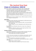
LEFT-LEFT = brain at rest
Basic metabolism (good balance between oxy and de-oxy Hb in bloodstream, normal usage of
oxygen by the neuronal cells, normal glucoses dosage as energy resource)
LEFT-RIGHT = activation of the neuron
→ Neurons will require an increased metabolism of glucoses and oxygen
Initially in bloodstream will be higher amount of de-oxy compared to oxy
→ This gives a reduced BOLD-signal
RIGHT = this triggers a haemodynamic respons rendering an increased transport of oxy to this region
of activation
Rendering in a lowering of the ratio de-oxy/oxy → rendering in an increase of BOLD-signals
3