Part 1: MRI – signal and contrast principles
Frequency and energy are proportional to each other → the more fotons = the higher the intensity
Introduction
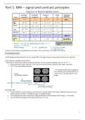
1H-MR images provide information → 1H = proton MRI = the signal we get is from protons (nucleus of an element)
proton density = hydrogen nuclei density
+ interactions of these nuclei with surrounding molecules (→ tissue specific relaxation times T1, T2, T2 * )
protons belong to water molecules. some have an evironment (surrounded by other water molecules)
T1 T2 T3 are typical MRI parameters
= T2 weighted image (water image) but
the ventricles who are filled with CF are
colours red so normally they are very
bright
Advantages MRI
• Provides different contrasts that you can measure to provide specific information. Different measures can be
obtained (proton density, T1-weighted image, T2 weighted image)
• Use radiofrequency waves, which have a high wavelength, but low frequency (low energy) – not harmful
• You can follow the progress of the disease because it is in vivo
1
, MRI image is 2D with pixel (picture element) that can be bright or
dark what wil be dependent on the parameter we bring into
contrast
What is the best parameter to see a difference?
3D had a voxel (volume element)
Free diffusion water nuclei → diffusion coefficient
tissue perfusion: (tumor, cerebralbloodflow)
Blood volume: blood flow
angiograms: visualization blood vessels(with/without contrast agent)
activated brain regions: functional MRI (fMRI)
Different imaging sequences provide:
• Anatomical information,
• physiological information
• functional information
• Molecular information, migration of labeled stem cells
few other nuclei may be used for specialised imaging purposes (e.g. 13C, 31P).
MRI is particularly suited to imaging differences between soft tissues, such as in the head, neck and spinal regions of the
body
CT is the technique to look at the bones --> in the brain it is used with contrast agents but with a healty brain there is not
much to see
Synopsis of MRI
Exposure of subject to big static magnetic field
0.2 (vet) → 0.5 T → 3 T (human) → 11.5 T (small animal)
(superconducting magnet –coil immersed in liquid He at 4K)
How can you generate a magnetic field? you need to coil. sent a certain potential to the coil
(rechterhand regel) it generates a horizontal magnetic field
electron magnet --> you can put it on and off
superconduction = what happens if you cool down the coil, the resistance will be decreased --> no resistance means that
the current going to the coil .... --> dangerous because some He will evaporate which increases the pressure --> fot safety
is there a pipe to evacuate the helium so you don't breath it in and also it is a huge magnet so you can't have any metal in
de room
1. Dedicated coil (part body): Transmit radio waves into subject [2~10 ms]
2. Turn off radio wave transmitter
3. Apply time-varying magnetic fields (position encoding)
4. Receive radio waves re-transmitted by subject
5. Convert measured RF data to image → you have to convert the raw data to an image
= RF coil, sends low energetic radiowaves after sending the body is emitting radio waves = MRI signal
2
,What kind of nuclei can be used for NMR?
Nucleus needs to have 2 properties:
• Spin
• charge
Nuclei are made of protons and neutrons
• Both have spin ½ → you have to have an odd number of spins
• Protons have charge
Pairs of spins tend to cancel, so only atoms with an odd number of protons or neutrons have spin
each proton has a magnetic momentum in a certain direction µ
Nuclear Magnetic Resonance: magnetic moment
spinning nucleus (1H) = positive charged sphere representing an electrical circular loop around the axis of rotation
Electrical current creates a magnetic field
Spin magnetic moment or spin (vector)
Angular momentum L (mass moving on circle velocity v →L=r x mv)
In an external magnetic field B0=B01z: Quantum mechanics: magnitude Lz restricted → z restricted
Most of the scanners have an horizontal field
proton has only 2 directions: spin up of parallel and spin down or antiparallel (against the direction of de field)
it can never be horizontal
3
, Motion of µ in B0-field
Spin system in external magnetic field B0
µ magnetic moment will experience a torque
torque = change in angular momentum → a force that creates a rotation (2e law of newton)
→ how will the vector µ change in time
➔ spin µ will precess around the magnetic field B0
➔ angular frequency of precession = Larmor frequency 0, proportional to strength
external magnetic field B0 :
Spin system in external magnetic field B0
Potential energy U, µ in B0-field
Interaction of magnetic moment µ in B0-field:
➔ Potential interaction energy:
➔ Zeeman interaction, split in 2 possible energy states
4
Frequency and energy are proportional to each other → the more fotons = the higher the intensity
Introduction
1H-MR images provide information → 1H = proton MRI = the signal we get is from protons (nucleus of an element)
proton density = hydrogen nuclei density
+ interactions of these nuclei with surrounding molecules (→ tissue specific relaxation times T1, T2, T2 * )
protons belong to water molecules. some have an evironment (surrounded by other water molecules)
T1 T2 T3 are typical MRI parameters
= T2 weighted image (water image) but
the ventricles who are filled with CF are
colours red so normally they are very
bright
Advantages MRI
• Provides different contrasts that you can measure to provide specific information. Different measures can be
obtained (proton density, T1-weighted image, T2 weighted image)
• Use radiofrequency waves, which have a high wavelength, but low frequency (low energy) – not harmful
• You can follow the progress of the disease because it is in vivo
1
, MRI image is 2D with pixel (picture element) that can be bright or
dark what wil be dependent on the parameter we bring into
contrast
What is the best parameter to see a difference?
3D had a voxel (volume element)
Free diffusion water nuclei → diffusion coefficient
tissue perfusion: (tumor, cerebralbloodflow)
Blood volume: blood flow
angiograms: visualization blood vessels(with/without contrast agent)
activated brain regions: functional MRI (fMRI)
Different imaging sequences provide:
• Anatomical information,
• physiological information
• functional information
• Molecular information, migration of labeled stem cells
few other nuclei may be used for specialised imaging purposes (e.g. 13C, 31P).
MRI is particularly suited to imaging differences between soft tissues, such as in the head, neck and spinal regions of the
body
CT is the technique to look at the bones --> in the brain it is used with contrast agents but with a healty brain there is not
much to see
Synopsis of MRI
Exposure of subject to big static magnetic field
0.2 (vet) → 0.5 T → 3 T (human) → 11.5 T (small animal)
(superconducting magnet –coil immersed in liquid He at 4K)
How can you generate a magnetic field? you need to coil. sent a certain potential to the coil
(rechterhand regel) it generates a horizontal magnetic field
electron magnet --> you can put it on and off
superconduction = what happens if you cool down the coil, the resistance will be decreased --> no resistance means that
the current going to the coil .... --> dangerous because some He will evaporate which increases the pressure --> fot safety
is there a pipe to evacuate the helium so you don't breath it in and also it is a huge magnet so you can't have any metal in
de room
1. Dedicated coil (part body): Transmit radio waves into subject [2~10 ms]
2. Turn off radio wave transmitter
3. Apply time-varying magnetic fields (position encoding)
4. Receive radio waves re-transmitted by subject
5. Convert measured RF data to image → you have to convert the raw data to an image
= RF coil, sends low energetic radiowaves after sending the body is emitting radio waves = MRI signal
2
,What kind of nuclei can be used for NMR?
Nucleus needs to have 2 properties:
• Spin
• charge
Nuclei are made of protons and neutrons
• Both have spin ½ → you have to have an odd number of spins
• Protons have charge
Pairs of spins tend to cancel, so only atoms with an odd number of protons or neutrons have spin
each proton has a magnetic momentum in a certain direction µ
Nuclear Magnetic Resonance: magnetic moment
spinning nucleus (1H) = positive charged sphere representing an electrical circular loop around the axis of rotation
Electrical current creates a magnetic field
Spin magnetic moment or spin (vector)
Angular momentum L (mass moving on circle velocity v →L=r x mv)
In an external magnetic field B0=B01z: Quantum mechanics: magnitude Lz restricted → z restricted
Most of the scanners have an horizontal field
proton has only 2 directions: spin up of parallel and spin down or antiparallel (against the direction of de field)
it can never be horizontal
3
, Motion of µ in B0-field
Spin system in external magnetic field B0
µ magnetic moment will experience a torque
torque = change in angular momentum → a force that creates a rotation (2e law of newton)
→ how will the vector µ change in time
➔ spin µ will precess around the magnetic field B0
➔ angular frequency of precession = Larmor frequency 0, proportional to strength
external magnetic field B0 :
Spin system in external magnetic field B0
Potential energy U, µ in B0-field
Interaction of magnetic moment µ in B0-field:
➔ Potential interaction energy:
➔ Zeeman interaction, split in 2 possible energy states
4