Summary MG: endocrine system and digestive and respiratory tract
Lecture 1 - Respiratory tract
Anatomy
Respiratory tract starts in the nose and mouth cavity.
- Humidify air.
- To facilitate the exchange of air at the blood-air interface in the alveoli (O2 is take up and
CO2 is released).
Nose: cavities conducting function, limited gas exchange.
Trachea: extra-pulmonary conducting function, limited gas exchange.
Thoracic cavity, bronchi and bronchioles conduction functions, limited gas exchange.
Alveoli: gas exchange.
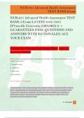
Large airways have airway smooth muscle which is connected between cartilage segments which
are shaped like a U (hoefijzer). Contraction of the smooth muscle
causes the cartilage to shrink which will lead to narrowing the lumen
of the large airways.
Small airways hereby the cartilage is present as loose fragments
and between these fragments there is smooth muscle. Also, when
these smooth muscles will contract there is a narrowing of the
lumen, but then of the small airways.
Upper airways: from the nose down to trachea and the main
bronchi.
- Epithelium: covers the airways, consists of:
o Ciliated cells.
o Mucous producing goblet cells.
- Submucosal gland (SMG): present in the upper airways,
produce mucous in response to neuronal stimulation by the
parasympathetic nervous system.
- Cartilage.
Small airways: bronchi, respiratory bronchioles and pleura with
lymphatic circulation.
- Cartilage: still present but now as islands.
- Glands: still there but much reduced.
- Epithelium: composition of the airway epithelium is different.
o Less involvement of the goblet cells.
- Morphology is the same as in the upper airways.
The further you go down you lose the density of glands and of mucous
producing goblet cells. So mucous production is mainly a feature in the
upper airways because at some point the smaller airways are so
small that obstruction will take place, which prevent you from
breathing.
Distal lung (alveoli): gas exchange will take place here.
Two types of cells:
- Alveolar epithelial cells: cover the alveoli.
, - Endothelial cells: make up pulmonary circulations.
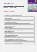
Airway epithelial cell heterogeneity:
- Ciliated cells: mucocilairy transport (clearance of the lungs from mucous) small motor
units that move mucous layer away from the lung into the esophagus.
o If this movement of mucous is not synchronized you have primary ciliary dyskinesia.
o More present in larger airways.
o Smoking causes destruction of the cilia, so people who smoke have shorter and less
dense cilia less muscularity clearance cough.
- Goblet cell / mucous cell: mucous production. Mucosa helps the airways protect against
particles or bacteria.
o More present in larger airways.
- Basal cells: airway progenitor (at the bottom of the epithelial cell layer). Repair the airways
when there is damage.
- Club cells: secretory proteins, small airway progenitor. Produce a more fluid secretion than
the goblet cells.
Mucociliairy transport
Alveolar epithelial cell heterogeneity
In the alveoli, all the previously mentioned epithelial cells are not present.
- Type 1 cells: O2/CO2 transport.
Flat thin cell that facilitates gas exchange (O2 and CO2) with neighboring pulmonary
microcirculation.
o Makes up 95% of the surface in the alveoli.
- Type 2 cells: surfactant, progenitor.
Bigger cells which have two functions:
o Produced surfactant which lowers the surface tensions make sure that the lung
will not collapse upon exhalation and is able to expand again upon inhalation.
Surfactant is produced relatively late during lung development (week 28). As
a result, prematurely born neonates require surfactant support in
combination with mechanical ventilation.
o Type 2 cells can replace the damaged type 1 cells. So, type 2 cells are progenitor cells
for type 1 cells.
Diseases of the respiratory tract
- Asthma.
- Allergic rhinitis.
- Cough.
- COPD.
- Pulmonary fibrosis.
- Cystic fibrosis.
Asthma
Definition: asthma is a heterogeneous disease, usually characterized by chronic airway inflammation.
It is defined by the history of respiratory symptoms such as wheeze, shortness of breath, chest
tightness and cough that vary over time and in intensity, together with variable expiratory airflow
limitation.
- Symptoms can be seasonal.
,Allergy and asthma
Majority (70%) of all asthma patients have allergic asthma.
- Non-allergic asthma is harder to treat.
Prevalence: allergies and asthma is the most common chronic disease, but less severe than other
chronic diseases like diabetes, cancer.
- Fetal cases have to do with lack of access to medicines.
- Rise in prevalence due to increased hygiene, we are less exposed to bacteria (live in cleaner
environment) immune system is responding to harmless elements.
Gender paradox: asthma is more prevalent in children than in adults, but children can grow out of it.
- Children asthma more common in males.
- Adults asthma more common in females, which can be explained in changes in hormones
females have a better immune system.
During puberty there is lung growth which is bigger for boys than for girls which may increase the
presence of asthma in women.
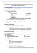
Airway hyperresponsiveness in asthma
Airway hyperresponsiveness in asthma: one of the main diagnostic
features used to diagnose asthma, this is a measured as change in so-
called FEV1.
Forced expiratory volume in 1 sec (FEV1): this is the amount of air than
you can exhale in 1 sec upon full inspiration.
When receiving a bronchoconstrictor: the FEV1 will drop because of
airflow obstruction by narrowing.
The higher the FEV1 number the more obstructive you will become. Healthy person will never
have a 20% drop.
So, if you did not reach a drop of 20% at a dose of 32 mg/mL, you are considered healthy.
In normal patients you see a plateau at the end of the curve. In asthma patients, the curve will not
reach a plateau but will keep increasing.
Airway hyperresponsiveness in asthma has its origin in a number of features
which are very specific for asthma.
- Genetic predisposition: can make you more suspected to asthma, but
not sufficient enough to develop asthma to develop asthma you
need subsequent exposure to environmental triggers.
- Transient airway hyperresponsiveness: mainly driven by
inflammation, which is usually the result of environmental triggers. A repeated exposure to
the triggers may result in airway remodeling.
- Permanent airway hyperresponsiveness: structural changes like a fibrotic process associated
with airway thickening.
Inflammation (allergic asthma, antibody response which drives the reaction)
Allergic asthma:
Antigen is picked up by an antigen presenting cell (dendritic cell or macrophage). The allergen is then
presented in the lymph node to naïve T-cells that will mature into CD4+ T-cells which can
differentiate into Th2 cells. These Th2 cells produce:
- IL-4: activate B-cells to produce IgE (mast cells are covered with these IgE antibodies, when
antigen meets this antibody histamine and protease release which causes
bronchoconstriction).
- IL-5 and IL-13: activate eosinophils (release protease and cytokines which will contribute to
mucous hyper secretion and oedema.
, This all will lead to airflow limitation.
Non allergic asthma:
Mast-cell response:
- For instance, cold air can facilitate the release of histamine from mast cell granules even in
absence of an allergen.
Alarmins: the pollutants to which the patient is exposed will cause micro injuries to the airway
epithelium. These injuries cause the epithelium to secrete cytokines that are also called alarmins: IL-
33, IL-25 and TSLP. These alarmins will activate the immune system and will activate innate lymphoid
cell type 2 (ILC2) cells which have similar functions to Th2 cells, like producing IL-5, IL-4 and IL-13.
This means that the end response is really similar (same symptoms) but the origin of the response
is different:
- Th2 important antigen in asthma.
- Alarmins important role in non-allergic asthma.
Glucocorticosteroids: used in asthma treatment to repress the inflammatory response.
- They inhibit cytokine production by Th2 cells.
- Antibodies can also be used against these cytokines to support the anti-inflammatory
treatment of asthma.
Mucous hypersecretion (persistent variations in the number of goblet cells), fibrosis of the airway
wall, and a rise in muscle mass all characteristics of asthma. The lumen will become constricted
and hypersensitive due to all of these characteristics.
Remodeling cannot yet be treated, only inflammation can be managed.
Airway structure (remodeling)
Mechanisms leading to remodeling are directly linked to inflammation and airway wall damage.
Repeated exposure to environmental substances will cause repeated injuries this will trigger the
inflammatory response and an attempt to airway repair.
- Normally when the cycle of airway healing is completed everything should be normal.
- When airway is not completely repaired or already re-injured during the repair cycle
activation the production of growth factors (TGFβ) and proteases which will cause
remodeling.
There have been no effective attempts to suppress the growth factors and therefore remodeling.
Because normal wound healing also depends on the same growth factors, which is harmful.
Allergen encounter – early and late response
The early and late response are typical for allergic asthma.
Lecture 1 - Respiratory tract
Anatomy
Respiratory tract starts in the nose and mouth cavity.
- Humidify air.
- To facilitate the exchange of air at the blood-air interface in the alveoli (O2 is take up and
CO2 is released).
Nose: cavities conducting function, limited gas exchange.
Trachea: extra-pulmonary conducting function, limited gas exchange.
Thoracic cavity, bronchi and bronchioles conduction functions, limited gas exchange.
Alveoli: gas exchange.
Large airways have airway smooth muscle which is connected between cartilage segments which
are shaped like a U (hoefijzer). Contraction of the smooth muscle
causes the cartilage to shrink which will lead to narrowing the lumen
of the large airways.
Small airways hereby the cartilage is present as loose fragments
and between these fragments there is smooth muscle. Also, when
these smooth muscles will contract there is a narrowing of the
lumen, but then of the small airways.
Upper airways: from the nose down to trachea and the main
bronchi.
- Epithelium: covers the airways, consists of:
o Ciliated cells.
o Mucous producing goblet cells.
- Submucosal gland (SMG): present in the upper airways,
produce mucous in response to neuronal stimulation by the
parasympathetic nervous system.
- Cartilage.
Small airways: bronchi, respiratory bronchioles and pleura with
lymphatic circulation.
- Cartilage: still present but now as islands.
- Glands: still there but much reduced.
- Epithelium: composition of the airway epithelium is different.
o Less involvement of the goblet cells.
- Morphology is the same as in the upper airways.
The further you go down you lose the density of glands and of mucous
producing goblet cells. So mucous production is mainly a feature in the
upper airways because at some point the smaller airways are so
small that obstruction will take place, which prevent you from
breathing.
Distal lung (alveoli): gas exchange will take place here.
Two types of cells:
- Alveolar epithelial cells: cover the alveoli.
, - Endothelial cells: make up pulmonary circulations.
Airway epithelial cell heterogeneity:
- Ciliated cells: mucocilairy transport (clearance of the lungs from mucous) small motor
units that move mucous layer away from the lung into the esophagus.
o If this movement of mucous is not synchronized you have primary ciliary dyskinesia.
o More present in larger airways.
o Smoking causes destruction of the cilia, so people who smoke have shorter and less
dense cilia less muscularity clearance cough.
- Goblet cell / mucous cell: mucous production. Mucosa helps the airways protect against
particles or bacteria.
o More present in larger airways.
- Basal cells: airway progenitor (at the bottom of the epithelial cell layer). Repair the airways
when there is damage.
- Club cells: secretory proteins, small airway progenitor. Produce a more fluid secretion than
the goblet cells.
Mucociliairy transport
Alveolar epithelial cell heterogeneity
In the alveoli, all the previously mentioned epithelial cells are not present.
- Type 1 cells: O2/CO2 transport.
Flat thin cell that facilitates gas exchange (O2 and CO2) with neighboring pulmonary
microcirculation.
o Makes up 95% of the surface in the alveoli.
- Type 2 cells: surfactant, progenitor.
Bigger cells which have two functions:
o Produced surfactant which lowers the surface tensions make sure that the lung
will not collapse upon exhalation and is able to expand again upon inhalation.
Surfactant is produced relatively late during lung development (week 28). As
a result, prematurely born neonates require surfactant support in
combination with mechanical ventilation.
o Type 2 cells can replace the damaged type 1 cells. So, type 2 cells are progenitor cells
for type 1 cells.
Diseases of the respiratory tract
- Asthma.
- Allergic rhinitis.
- Cough.
- COPD.
- Pulmonary fibrosis.
- Cystic fibrosis.
Asthma
Definition: asthma is a heterogeneous disease, usually characterized by chronic airway inflammation.
It is defined by the history of respiratory symptoms such as wheeze, shortness of breath, chest
tightness and cough that vary over time and in intensity, together with variable expiratory airflow
limitation.
- Symptoms can be seasonal.
,Allergy and asthma
Majority (70%) of all asthma patients have allergic asthma.
- Non-allergic asthma is harder to treat.
Prevalence: allergies and asthma is the most common chronic disease, but less severe than other
chronic diseases like diabetes, cancer.
- Fetal cases have to do with lack of access to medicines.
- Rise in prevalence due to increased hygiene, we are less exposed to bacteria (live in cleaner
environment) immune system is responding to harmless elements.
Gender paradox: asthma is more prevalent in children than in adults, but children can grow out of it.
- Children asthma more common in males.
- Adults asthma more common in females, which can be explained in changes in hormones
females have a better immune system.
During puberty there is lung growth which is bigger for boys than for girls which may increase the
presence of asthma in women.
Airway hyperresponsiveness in asthma
Airway hyperresponsiveness in asthma: one of the main diagnostic
features used to diagnose asthma, this is a measured as change in so-
called FEV1.
Forced expiratory volume in 1 sec (FEV1): this is the amount of air than
you can exhale in 1 sec upon full inspiration.
When receiving a bronchoconstrictor: the FEV1 will drop because of
airflow obstruction by narrowing.
The higher the FEV1 number the more obstructive you will become. Healthy person will never
have a 20% drop.
So, if you did not reach a drop of 20% at a dose of 32 mg/mL, you are considered healthy.
In normal patients you see a plateau at the end of the curve. In asthma patients, the curve will not
reach a plateau but will keep increasing.
Airway hyperresponsiveness in asthma has its origin in a number of features
which are very specific for asthma.
- Genetic predisposition: can make you more suspected to asthma, but
not sufficient enough to develop asthma to develop asthma you
need subsequent exposure to environmental triggers.
- Transient airway hyperresponsiveness: mainly driven by
inflammation, which is usually the result of environmental triggers. A repeated exposure to
the triggers may result in airway remodeling.
- Permanent airway hyperresponsiveness: structural changes like a fibrotic process associated
with airway thickening.
Inflammation (allergic asthma, antibody response which drives the reaction)
Allergic asthma:
Antigen is picked up by an antigen presenting cell (dendritic cell or macrophage). The allergen is then
presented in the lymph node to naïve T-cells that will mature into CD4+ T-cells which can
differentiate into Th2 cells. These Th2 cells produce:
- IL-4: activate B-cells to produce IgE (mast cells are covered with these IgE antibodies, when
antigen meets this antibody histamine and protease release which causes
bronchoconstriction).
- IL-5 and IL-13: activate eosinophils (release protease and cytokines which will contribute to
mucous hyper secretion and oedema.
, This all will lead to airflow limitation.
Non allergic asthma:
Mast-cell response:
- For instance, cold air can facilitate the release of histamine from mast cell granules even in
absence of an allergen.
Alarmins: the pollutants to which the patient is exposed will cause micro injuries to the airway
epithelium. These injuries cause the epithelium to secrete cytokines that are also called alarmins: IL-
33, IL-25 and TSLP. These alarmins will activate the immune system and will activate innate lymphoid
cell type 2 (ILC2) cells which have similar functions to Th2 cells, like producing IL-5, IL-4 and IL-13.
This means that the end response is really similar (same symptoms) but the origin of the response
is different:
- Th2 important antigen in asthma.
- Alarmins important role in non-allergic asthma.
Glucocorticosteroids: used in asthma treatment to repress the inflammatory response.
- They inhibit cytokine production by Th2 cells.
- Antibodies can also be used against these cytokines to support the anti-inflammatory
treatment of asthma.
Mucous hypersecretion (persistent variations in the number of goblet cells), fibrosis of the airway
wall, and a rise in muscle mass all characteristics of asthma. The lumen will become constricted
and hypersensitive due to all of these characteristics.
Remodeling cannot yet be treated, only inflammation can be managed.
Airway structure (remodeling)
Mechanisms leading to remodeling are directly linked to inflammation and airway wall damage.
Repeated exposure to environmental substances will cause repeated injuries this will trigger the
inflammatory response and an attempt to airway repair.
- Normally when the cycle of airway healing is completed everything should be normal.
- When airway is not completely repaired or already re-injured during the repair cycle
activation the production of growth factors (TGFβ) and proteases which will cause
remodeling.
There have been no effective attempts to suppress the growth factors and therefore remodeling.
Because normal wound healing also depends on the same growth factors, which is harmful.
Allergen encounter – early and late response
The early and late response are typical for allergic asthma.