The Body's Purifiers: A Detailed Exploration of the
Human Excretory and Urinary System
The human excretory system, often specifically referred to as the urinary system due to the
kidneys' central role, is a vital biological system dedicated to filtering waste products from the
blood and expelling them from the body. Beyond mere waste removal, this sophisticated system
plays an indispensable role in maintaining the body's internal environment (homeostasis) by
regulating blood volume, blood pressure, electrolyte levels, and acid-base balance. Without its
continuous operation, toxic substances would accumulate, rapidly leading to severe health
complications.
I. Components of the Urinary System:
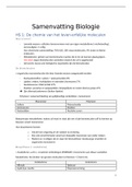
The urinary system consists of a pair of kidneys, two ureters, a urinary bladder, and a urethra.
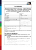
1. Kidneys:
○ Location: Two bean-shaped organs located on either side of the spine, just
below the rib cage, behind the abdominal peritoneum. The right kidney is typically
slightly lower than the left due to the liver's position.
○ Function: The primary blood-filtering organs. They process about 180 liters of
blood plasma daily, producing roughly 1-2 liters of urine.
○ Structure:
■ Renal Cortex: The outermost layer, granular in appearance, containing
the renal corpuscles and convoluted tubules of the nephrons.
■ Renal Medulla: The inner region, composed of cone-shaped structures
called renal pyramids. These pyramids contain the loops of Henle and
collecting ducts.
■ Renal Pelvis: A funnel-shaped structure in the center of the kidney that
collects urine from the collecting ducts and funnels it into the ureter.
2. Ureters:
○ Two slender, muscular tubes (about 25-30 cm long) that extend from the renal
pelvis of each kidney to the posterior wall of the urinary bladder.
○ Function: They transport urine from the kidneys to the bladder through peristaltic
contractions of their smooth muscle walls.
3. Urinary Bladder:
○ A hollow, muscular, distensible organ located in the pelvic cavity, posterior to the
pubic symphysis.
○ Function: Serves as a temporary storage reservoir for urine. Its muscular walls
(detrusor muscle) contract during urination (micturition) to expel urine.
○ Capacity: Can typically hold 400-600 ml of urine, though the urge to urinate
usually begins around 200-300 ml.
4. Urethra:
, ○ A tube that extends from the floor of the urinary bladder to the outside of the
body.
○ Function: Serves as the exit pathway for urine.
○ Differences (Male vs. Female):
■ Female Urethra: Shorter (about 3-4 cm) and serves only the urinary
function.
■ Male Urethra: Longer (about 15-20 cm) and serves both urinary and
reproductive (ejaculation of semen) functions.
○ Sphincters: Both internal (involuntary smooth muscle) and external (voluntary
skeletal muscle) urethral sphincters control the flow of urine.
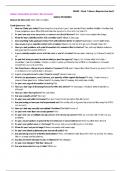
II. The Nephron: The Functional Unit of the Kidney:
Each kidney contains over a million microscopic filtering units called nephrons. The nephron is
the fundamental structural and functional unit responsible for forming urine.
1. Components of a Nephron:
○ Renal Corpuscle: The filtering component.
■ Glomerulus: A tuft of capillaries where blood filtration begins.
■ Bowman's (Glomerular) Capsule: A double-walled cup that surrounds
the glomerulus and collects the filtered fluid (filtrate).
○ Renal Tubule: A long, convoluted tube extending from Bowman's capsule.
■ Proximal Convoluted Tubule (PCT): Highly coiled, responsible for most
reabsorption of water, ions, and organic nutrients.
■ Loop of Henle: A U-shaped segment that extends into the renal medulla,
crucial for establishing the osmotic gradient necessary for concentrating
urine. It has descending and ascending limbs.
■ Distal Convoluted Tubule (DCT): Coiled, involved in fine-tuning
reabsorption and secretion, particularly under hormonal control.
■ Collecting Duct: Receives filtrate from multiple DCTs and extends
through the renal medulla, playing a key role in final urine concentration.
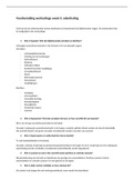
III. Physiology of Urine Formation: A Three-Step Process:
Urine formation involves a continuous, three-step process within the nephrons:
1. Glomerular Filtration:
○ Location: Occurs in the renal corpuscle (glomerulus and Bowman's capsule).
○ Process: Blood enters the glomerulus under high pressure. The thin, porous
walls of the glomerular capillaries act as a filter, forcing water and small solutes
(ions, glucose, amino acids, urea, creatinine) from the blood into Bowman's
capsule, forming the glomerular filtrate.
○ Exclusions: Blood cells, large proteins, and other large molecules are typically
too large to pass through the filter and remain in the blood.
2. Tubular Reabsorption:
Human Excretory and Urinary System
The human excretory system, often specifically referred to as the urinary system due to the
kidneys' central role, is a vital biological system dedicated to filtering waste products from the
blood and expelling them from the body. Beyond mere waste removal, this sophisticated system
plays an indispensable role in maintaining the body's internal environment (homeostasis) by
regulating blood volume, blood pressure, electrolyte levels, and acid-base balance. Without its
continuous operation, toxic substances would accumulate, rapidly leading to severe health
complications.
I. Components of the Urinary System:
The urinary system consists of a pair of kidneys, two ureters, a urinary bladder, and a urethra.
1. Kidneys:
○ Location: Two bean-shaped organs located on either side of the spine, just
below the rib cage, behind the abdominal peritoneum. The right kidney is typically
slightly lower than the left due to the liver's position.
○ Function: The primary blood-filtering organs. They process about 180 liters of
blood plasma daily, producing roughly 1-2 liters of urine.
○ Structure:
■ Renal Cortex: The outermost layer, granular in appearance, containing
the renal corpuscles and convoluted tubules of the nephrons.
■ Renal Medulla: The inner region, composed of cone-shaped structures
called renal pyramids. These pyramids contain the loops of Henle and
collecting ducts.
■ Renal Pelvis: A funnel-shaped structure in the center of the kidney that
collects urine from the collecting ducts and funnels it into the ureter.
2. Ureters:
○ Two slender, muscular tubes (about 25-30 cm long) that extend from the renal
pelvis of each kidney to the posterior wall of the urinary bladder.
○ Function: They transport urine from the kidneys to the bladder through peristaltic
contractions of their smooth muscle walls.
3. Urinary Bladder:
○ A hollow, muscular, distensible organ located in the pelvic cavity, posterior to the
pubic symphysis.
○ Function: Serves as a temporary storage reservoir for urine. Its muscular walls
(detrusor muscle) contract during urination (micturition) to expel urine.
○ Capacity: Can typically hold 400-600 ml of urine, though the urge to urinate
usually begins around 200-300 ml.
4. Urethra:
, ○ A tube that extends from the floor of the urinary bladder to the outside of the
body.
○ Function: Serves as the exit pathway for urine.
○ Differences (Male vs. Female):
■ Female Urethra: Shorter (about 3-4 cm) and serves only the urinary
function.
■ Male Urethra: Longer (about 15-20 cm) and serves both urinary and
reproductive (ejaculation of semen) functions.
○ Sphincters: Both internal (involuntary smooth muscle) and external (voluntary
skeletal muscle) urethral sphincters control the flow of urine.
II. The Nephron: The Functional Unit of the Kidney:
Each kidney contains over a million microscopic filtering units called nephrons. The nephron is
the fundamental structural and functional unit responsible for forming urine.
1. Components of a Nephron:
○ Renal Corpuscle: The filtering component.
■ Glomerulus: A tuft of capillaries where blood filtration begins.
■ Bowman's (Glomerular) Capsule: A double-walled cup that surrounds
the glomerulus and collects the filtered fluid (filtrate).
○ Renal Tubule: A long, convoluted tube extending from Bowman's capsule.
■ Proximal Convoluted Tubule (PCT): Highly coiled, responsible for most
reabsorption of water, ions, and organic nutrients.
■ Loop of Henle: A U-shaped segment that extends into the renal medulla,
crucial for establishing the osmotic gradient necessary for concentrating
urine. It has descending and ascending limbs.
■ Distal Convoluted Tubule (DCT): Coiled, involved in fine-tuning
reabsorption and secretion, particularly under hormonal control.
■ Collecting Duct: Receives filtrate from multiple DCTs and extends
through the renal medulla, playing a key role in final urine concentration.
III. Physiology of Urine Formation: A Three-Step Process:
Urine formation involves a continuous, three-step process within the nephrons:
1. Glomerular Filtration:
○ Location: Occurs in the renal corpuscle (glomerulus and Bowman's capsule).
○ Process: Blood enters the glomerulus under high pressure. The thin, porous
walls of the glomerular capillaries act as a filter, forcing water and small solutes
(ions, glucose, amino acids, urea, creatinine) from the blood into Bowman's
capsule, forming the glomerular filtrate.
○ Exclusions: Blood cells, large proteins, and other large molecules are typically
too large to pass through the filter and remain in the blood.
2. Tubular Reabsorption: