Samenvatting Developmental Biology
H1: Vertebrate development
1.1: Development
1.1.1: Frog development
Typical model organism = Xenopus laevis from order Anura = African clawed frog => aquatic carnivorous frog.
In experiments:
• Injection of hormones (hCG) to induce egg laying
• In vitro fertilization of egg
Development speed depends on temperature
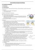
Egg = frog oocyte (up to 1mm in diameter), has 2 hemispheres:
• Animal hemisphere (dark pigmented)
• Vegetal hemisphere (light pigmented) => there you can find yolk platelets (food during
development), which is heavy. This causes orientation of oocyte due to gravity & has a
slower division than the rest.
=> These 2 hemispheres define the animal-vegetal axis.
Development:
1. The sperm entry point is on ventral side (= left top side & opposite to Nieuwkoop
center). Fertilization happens in animal hemisphere.
2. Membrane changes so that no further sperm can enter.
3. After 30 minutes the cytoskeleton makes the cortex rotates 30° towards sperm
entry point. This is a visible change due to pigment in animal hemisphere. During
rotation gravity keeps oocyte in same place. Cortical rotation is critical for original
asymmetry of embryo.
4. Cleavage = mitosis process of dividing cells. During cleavage the volume of frog cell stays the same.
o 1st cleavage => is a vertical cleavage that starts from animal pole straight to vegetal pole. Now you have
2 blastomeres (= left & right side of embryo).
o 2nd cleavage => is a vertical cleavage at right angle with first one & starts before first cleavage is finished,
but is slowed down by heavy yolk.
o 3rd cleavage => is a horizontal cleavage & more in the animal hemisphere due to yolk. Now you have 4
small blastomeres in animal hemisphere & 4 big ones in vegetal hemisphere.
o Synchronous cleavages => leads to lots of small cells in animal hemisphere & a small number of large yolk
cells in vegetal hemisphere
5. Blastula at 128 cell stage => here you have formation of blastocoel (= cavity filled with fluid at
animal region & is derived from small intracellular space at 2 cell stage). Blastocoel functions
as a migration space for gastrulating cells & isolation of animal cap from inductive signals
from vegetal region. All divisions until blastula stage happen extremely fast, with S (=
chromosome duplications) & M (= mitosis) phase follow each other & absence of G1 & G2
phase. This gives cycles of 30 minutes instead of 16h. no transcription, as all proteins &
mRNA are derived from maternal genome.
6. After 12 divisions cycles = mid blastula transition (MBT) => start of zygotic transcription.
7. Gastrulation (see 1.2)
8. Neurulation
I. Mesoderm forms Notochord (= rod-like structure in midline under ectoderm).
II. Ectoderm above notochord thickens into neural plate.
III. Neural plate folds into a groove, then the edges (= neural folds) rise & fuse = Neural tube (will become
brain & spinal cord).
, IV. Neural crest cells (from neural plate edges) break off & migrate throughout body & they will form parts of
the PNS, pigment cells, bones of the face, etc.
V. Mesoderm next to notochord organizes into somites (give rise to muscles, vertebrae, dermis of skin).
9. Organogenesis => Each germ layer (ectoderm, mesoderm, endoderm) starts forming specific organs & tissues,
this leads to the formation of a typical tadpole.
10. Hatching of tadpole
1.1.2: Mammalian Development
Typical model organism for genetic tool is the mouse.
A mouse has 2 uteri & 8 to 12 embryo’s per female. Their eggs are 100 microns in diameter. The use of an inbred line
will give no genetic variation between the mice.
Development:
1. Sperm plug is formed 0.5 days post coitum (dpc).
2. After fertilization in the oviduct (humans is in Fallopian tube), the egg has now 2 pronuclei & a zona pelicula.
3. Cleavage => Cell division is not synchronous (different from frog).
o 1st cleavage happens after 24 h = normal meridional division
o 2nd cleavage happens after 12 h = 1 blastomere divides meridional & another equatorial
4. Zygotic transcription starts between 1 & 8 cell stage
5. At 8 cell stage there is compaction = loss of morula shape & ball
formation with strong adherence of cells. Outside membranes
have numerous microvilli, while membranes that are in contact
with other cells are smooth (= Polarization of cells). Cells no
longer totipotent (before compaction each cell can give rise to a
complete embryo).
6. At 16 cell stage tight junctions between outside cells are formed
& gap junction between inside cells.
7. Formation of blastocyst at 64 cells stage by pumping ions between cells (change in osmolality allows passive
water movement). It has 3 layers:
o Trophectoderm (TE) = later extraembryonic tissue (parietal yolk sack). They were polarized cells during
compaction.
o Primitive endoderm (PrEnd) = later extraembryonic tissue (visceral yolk sack) .
o Inner cell mass (ICM) = later give rise to adult & some extraembryonic tissue (protected by TE & PrEnd
from outside) = 13 cells of 64 .
8. Blastocyst reaches uterus & hatches (free of zona pellucida). There is implantation, meaning that trophoblast
cells from embryo & uterine cells proliferate. These will surround embryo.
9. After implantation, the inner cell mass differentiates into epiblasts.
10. Epiblast begins to grow & expand, forming around 1000 cells. Proamniotic cavity form within expanding
epiblast. Epiblast forms cup-shaped structure around the cavity & embryo has morphology = egg cylinder
11. Primitive endoderm (from ICM) spread along the inner surface of trophectoderm & differentiates into visceral
endoderm (adjacent to epiblast) parietal endoderm (adjacent to trophectoderm). Up until here the embryo has
radial symmetry => impossible to distinguish anterior from posterior
12. Gastrulation (see 1.2)
13. Neurulation
I. Out of node, the notochord & endoderm will derive. Embryo is then composed of 3 germ layers.
II. Mesoderm on both sides of neural tube form somites.
III. Neural tube flanked by somites is forming & is positioned above the notochord.
IV. Invagination under neural fold = foregut pocket.
V. Turning of embryo in “classical” foetal position & wrap embryo in extraembryonic membranes.
14. Organogenesis
, 1.2: Gastrulation Frog VS Mouse
Gastrulation frog:
1. Gastrulation starts at 180° from the sperm entry point (= future dorsal side).
2. Bottle cells form here by apical constriction & elongation. They form a small groove in embryo dorsal blastopore
lip, the opening itself is the blastopore. By convention, the forming blastopore lip is shown on lower right side.
3. Involution at blastopore lip = sheets of cells roll inward & migrates toward the animal pole. These internalized
cells are dorsal mesoderm precursors (notochord, somites).
4. At animal pole they form the archenteron (= future gut).
5. Blastopore lip extends laterally & ventrally forming a ring around vegetal hemisphere. Cells entering through
will form
o Dorsal lip => notochord & paraxial mesoderm
o Lateral lip => intermediate mesoderm
o Ventral lip => blood, kidney, lateral plate mesoderm
6. Ectodermal cells spread over embryo by epiboly & intercalation.
7. Dorsal mesoderm undergoes convergence-extension which results in antero-posterior elongation & this is
essential for body axis extension.
8. The archenteron continuous to expand & replace the blastocoel.
9. Blastopore closes progressively & at this site of closure, the remaining vegetal yolk cells form yulk plug.
10. By the end you have
o Ectoderm on the outside => will form epidermis & neural tissue
o Mesoderm in the middle => will form muscle, bone, connective tissue, blood, gonads & kidney
o Endoderm on the inside => will form gut lining & internal organs
3 cellular behaviors leading to changes in shape of cell layer:
• Epiboly = increase of surface resulting from a change in cell shape. The cells become thinner & spread more, it
can occur in 1-cell thick sheets. This movement is typical of external cell layer of gastrula (ectoderm).
• Intercalation = process during which several cell layers mix with one another resulting in thinner but more
extensive sheet of cells. Typical for mesoderm.
• Convergence-Extension = specialized case of intercalation leading to highly directional spreading of the cell
sheet. It is composed of 2 intercalation movements.
o Radial intercalation = Cells from 2 different layers mix, resulting in spreading of the sheet.
o Medio-lateral intercalation = Cells from same sheet intercalate in a particular direction & this causes a
directional spreading.
Gastrulation mouse:
1. Start gastrulation with appearance of a primitive streak forming a thickening at junction between embryonic &
extraembryonic epiblast.
2. Epiblast cells at junction between embryonic & extraembryonic undergo epithelial-to-mesenchymal transition
(EMT), delamination & ingress into primitive streak.
H1: Vertebrate development
1.1: Development
1.1.1: Frog development
Typical model organism = Xenopus laevis from order Anura = African clawed frog => aquatic carnivorous frog.
In experiments:
• Injection of hormones (hCG) to induce egg laying
• In vitro fertilization of egg
Development speed depends on temperature
Egg = frog oocyte (up to 1mm in diameter), has 2 hemispheres:
• Animal hemisphere (dark pigmented)
• Vegetal hemisphere (light pigmented) => there you can find yolk platelets (food during
development), which is heavy. This causes orientation of oocyte due to gravity & has a
slower division than the rest.
=> These 2 hemispheres define the animal-vegetal axis.
Development:
1. The sperm entry point is on ventral side (= left top side & opposite to Nieuwkoop
center). Fertilization happens in animal hemisphere.
2. Membrane changes so that no further sperm can enter.
3. After 30 minutes the cytoskeleton makes the cortex rotates 30° towards sperm
entry point. This is a visible change due to pigment in animal hemisphere. During
rotation gravity keeps oocyte in same place. Cortical rotation is critical for original
asymmetry of embryo.
4. Cleavage = mitosis process of dividing cells. During cleavage the volume of frog cell stays the same.
o 1st cleavage => is a vertical cleavage that starts from animal pole straight to vegetal pole. Now you have
2 blastomeres (= left & right side of embryo).
o 2nd cleavage => is a vertical cleavage at right angle with first one & starts before first cleavage is finished,
but is slowed down by heavy yolk.
o 3rd cleavage => is a horizontal cleavage & more in the animal hemisphere due to yolk. Now you have 4
small blastomeres in animal hemisphere & 4 big ones in vegetal hemisphere.
o Synchronous cleavages => leads to lots of small cells in animal hemisphere & a small number of large yolk
cells in vegetal hemisphere
5. Blastula at 128 cell stage => here you have formation of blastocoel (= cavity filled with fluid at
animal region & is derived from small intracellular space at 2 cell stage). Blastocoel functions
as a migration space for gastrulating cells & isolation of animal cap from inductive signals
from vegetal region. All divisions until blastula stage happen extremely fast, with S (=
chromosome duplications) & M (= mitosis) phase follow each other & absence of G1 & G2
phase. This gives cycles of 30 minutes instead of 16h. no transcription, as all proteins &
mRNA are derived from maternal genome.
6. After 12 divisions cycles = mid blastula transition (MBT) => start of zygotic transcription.
7. Gastrulation (see 1.2)
8. Neurulation
I. Mesoderm forms Notochord (= rod-like structure in midline under ectoderm).
II. Ectoderm above notochord thickens into neural plate.
III. Neural plate folds into a groove, then the edges (= neural folds) rise & fuse = Neural tube (will become
brain & spinal cord).
, IV. Neural crest cells (from neural plate edges) break off & migrate throughout body & they will form parts of
the PNS, pigment cells, bones of the face, etc.
V. Mesoderm next to notochord organizes into somites (give rise to muscles, vertebrae, dermis of skin).
9. Organogenesis => Each germ layer (ectoderm, mesoderm, endoderm) starts forming specific organs & tissues,
this leads to the formation of a typical tadpole.
10. Hatching of tadpole
1.1.2: Mammalian Development
Typical model organism for genetic tool is the mouse.
A mouse has 2 uteri & 8 to 12 embryo’s per female. Their eggs are 100 microns in diameter. The use of an inbred line
will give no genetic variation between the mice.
Development:
1. Sperm plug is formed 0.5 days post coitum (dpc).
2. After fertilization in the oviduct (humans is in Fallopian tube), the egg has now 2 pronuclei & a zona pelicula.
3. Cleavage => Cell division is not synchronous (different from frog).
o 1st cleavage happens after 24 h = normal meridional division
o 2nd cleavage happens after 12 h = 1 blastomere divides meridional & another equatorial
4. Zygotic transcription starts between 1 & 8 cell stage
5. At 8 cell stage there is compaction = loss of morula shape & ball
formation with strong adherence of cells. Outside membranes
have numerous microvilli, while membranes that are in contact
with other cells are smooth (= Polarization of cells). Cells no
longer totipotent (before compaction each cell can give rise to a
complete embryo).
6. At 16 cell stage tight junctions between outside cells are formed
& gap junction between inside cells.
7. Formation of blastocyst at 64 cells stage by pumping ions between cells (change in osmolality allows passive
water movement). It has 3 layers:
o Trophectoderm (TE) = later extraembryonic tissue (parietal yolk sack). They were polarized cells during
compaction.
o Primitive endoderm (PrEnd) = later extraembryonic tissue (visceral yolk sack) .
o Inner cell mass (ICM) = later give rise to adult & some extraembryonic tissue (protected by TE & PrEnd
from outside) = 13 cells of 64 .
8. Blastocyst reaches uterus & hatches (free of zona pellucida). There is implantation, meaning that trophoblast
cells from embryo & uterine cells proliferate. These will surround embryo.
9. After implantation, the inner cell mass differentiates into epiblasts.
10. Epiblast begins to grow & expand, forming around 1000 cells. Proamniotic cavity form within expanding
epiblast. Epiblast forms cup-shaped structure around the cavity & embryo has morphology = egg cylinder
11. Primitive endoderm (from ICM) spread along the inner surface of trophectoderm & differentiates into visceral
endoderm (adjacent to epiblast) parietal endoderm (adjacent to trophectoderm). Up until here the embryo has
radial symmetry => impossible to distinguish anterior from posterior
12. Gastrulation (see 1.2)
13. Neurulation
I. Out of node, the notochord & endoderm will derive. Embryo is then composed of 3 germ layers.
II. Mesoderm on both sides of neural tube form somites.
III. Neural tube flanked by somites is forming & is positioned above the notochord.
IV. Invagination under neural fold = foregut pocket.
V. Turning of embryo in “classical” foetal position & wrap embryo in extraembryonic membranes.
14. Organogenesis
, 1.2: Gastrulation Frog VS Mouse
Gastrulation frog:
1. Gastrulation starts at 180° from the sperm entry point (= future dorsal side).
2. Bottle cells form here by apical constriction & elongation. They form a small groove in embryo dorsal blastopore
lip, the opening itself is the blastopore. By convention, the forming blastopore lip is shown on lower right side.
3. Involution at blastopore lip = sheets of cells roll inward & migrates toward the animal pole. These internalized
cells are dorsal mesoderm precursors (notochord, somites).
4. At animal pole they form the archenteron (= future gut).
5. Blastopore lip extends laterally & ventrally forming a ring around vegetal hemisphere. Cells entering through
will form
o Dorsal lip => notochord & paraxial mesoderm
o Lateral lip => intermediate mesoderm
o Ventral lip => blood, kidney, lateral plate mesoderm
6. Ectodermal cells spread over embryo by epiboly & intercalation.
7. Dorsal mesoderm undergoes convergence-extension which results in antero-posterior elongation & this is
essential for body axis extension.
8. The archenteron continuous to expand & replace the blastocoel.
9. Blastopore closes progressively & at this site of closure, the remaining vegetal yolk cells form yulk plug.
10. By the end you have
o Ectoderm on the outside => will form epidermis & neural tissue
o Mesoderm in the middle => will form muscle, bone, connective tissue, blood, gonads & kidney
o Endoderm on the inside => will form gut lining & internal organs
3 cellular behaviors leading to changes in shape of cell layer:
• Epiboly = increase of surface resulting from a change in cell shape. The cells become thinner & spread more, it
can occur in 1-cell thick sheets. This movement is typical of external cell layer of gastrula (ectoderm).
• Intercalation = process during which several cell layers mix with one another resulting in thinner but more
extensive sheet of cells. Typical for mesoderm.
• Convergence-Extension = specialized case of intercalation leading to highly directional spreading of the cell
sheet. It is composed of 2 intercalation movements.
o Radial intercalation = Cells from 2 different layers mix, resulting in spreading of the sheet.
o Medio-lateral intercalation = Cells from same sheet intercalate in a particular direction & this causes a
directional spreading.
Gastrulation mouse:
1. Start gastrulation with appearance of a primitive streak forming a thickening at junction between embryonic &
extraembryonic epiblast.
2. Epiblast cells at junction between embryonic & extraembryonic undergo epithelial-to-mesenchymal transition
(EMT), delamination & ingress into primitive streak.