Technieken
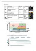
Full name ‘Waves’ Processes Image
CT Radiography X-ray Ana –
Computerized Tomography (Phys)
MRI Magnetic Resonance Radiowave Ana – Phys
Imaging s - Meta–
(Mole)
PET Positron Emission γ-wave (Phys) –
SPECT Tomography Meta - Mole
Single Photon CT
Optic Optical imaging Visible light Mole –
al Bioluminescence imaging meta –
(phys)
US UltraSound Soundwave Ana – Phys
(echo) s – (Meta)
,MRI
General:
Magnet
Superconducting coils
o Shim coils
o RF coils
o Gradient coils
Magnetic field (M)
o Has Field strength (B) in Tesla (T)
Clinical: 0,5-3T resolution: 1-3mm
Preclinical: 7-9,3T resolution: 70-125µm
o Generated by coils
o Higer field higher resolution (better alignment of + protons in
M)
NMR (= Nuclear Magnetic Resonance)
o Signal from protons (Hydrogen)!! has magnetic moment: ½ spin
electric current Mz in time
o Alignment towards M
Parallel (low energy state) OR anti-parallel
Precision = protons never perfectly aligned with M
Protons at equilibrium (Mz) Give F pulse (Mxy) change
orientation restore over time T1 or T2
Relaxation processes Gives contrast
o T1 – relaxation (restored to normal Mz)
o T2 – relaxation (not restored 0)
TR / TE time Image Liquid Fat
(ms)
T1 Short (500/15) Brighter Dark Bright
T2 Long (3000/90) Darker Bright Dark
, Structural Measurement
Volumetric MRI
o Spatial Normalization
o Huntington Disease
Contrast agents
o Diamagnetic ions only paired e- antiparallel to B0
o Paramagnetic ions one unpaired e- parallel to B0 shorten
T1 of water
Gadolinium leakage in BBB
o Ferromagnetic ions dephasing faster (T2) darker contrast
enhanced
Fe2+ ( vb: to detect Microbleeds)
Diffusion microstructural MRI
o Isotropic diffusion profile (low FA) Free diffusion
o
o Anisotropic diffusion profile (high FA) Preference diffusion
Axon has preference diffusion (λ1 forward)
λ1 = Axial Diffusity (AD)
λ2 , λ3 = Radial Diffusity
(RD)
o