Task 1:
Anatomy of the brain
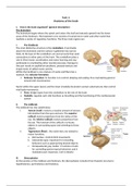
1. How is the brain organized? (general description)
The brainstem
The brainstem begins where the spinal cord enters the skull and extends upward into the lower
areas of the forebrain. The brainstem core consists of cranial-nerve nuclei and other nuclei that
mediate a variety of regulatory functions. The three main regions are:
i. The hindbrain
The most distinctive structure is the cerebellum. It protrudes
above the brainstem and the surface is gathered into narrow
folds. At the base of the cerebellum are several nuclei that send
connections to other parts of the brain. The cerebellum plays a
role in (fine) motor coordination and motor learning and may
participate in coordinating other mental processes. Damage to
this part results in equilibrium problems, postural defects and
impairments of skilled motor activity.
Within the hindbrain’s core mixture of nuclei and fibers lies a
network, the reticular formation.
Reticular formation: its function is to control sleeping and waking, thus maintaining general
arousal and consciousness.
Nuclei within the upper (pons) and the lower (medulla) brainstem contain substructures that control
vital body movements:
Pons: bridge inputs from the cerebellum to the rest of the brain
Medulla: regulate such vital functions as breathing and the functioning of the cardiovascular
system
ii. The midbrain
The midbrain has two subdivisions:
i. Tectum (roof): receives a massive amount of sensory
information from the eyes and ears. The superior
colliculi receive projections from the retina of the
eye, the inferior colliculi receives projections from
the ear. The function of the colliculi is to locate
objects in surrounding space and orienting to those
objects.
ii. Tegmentum (floor): the nuclei here are related to
motor functions.
a. Red nucleus: controls limb movements
b. Substantia nigra: important for rewarding
behaviors such as approaching desired objects
c. Periaqueductal gray matter: it contains circuits
for controlling species-typical behaviors and it
modulates pain responses. (not in details)
iii. Diencephalon
At the junction of the midbrain and forebrain, the diencephalon includes three thalamic structures:
hypothalamus, epi-thalamus and thalamus.
, Hypothalamus: connects to and interacts with the pituitary gland to control many endocrine
functions. It allows us to respond to the internal and external environment and to maintain
homeostasis. The hypothalamus is structurally part of the diencephalon but it functions as
part of the limbic system.
Epi-thalamus: it is a collection of nuclei at the posterior of the diencephalon. It secretes the
hormone melatonin, which influences daily and seasonal body rhythms.
Thalamus: it is a nucleus that serves as a hub interconnecting many brain regions. All the
information the cortex receives, is first relayed through the thalamus.
o One group of thalamic nuclei relays information from sensory systems to their
appropriate targets.
The lateral geniculate body receives visual projections The medial geniculate
body receives auditory projections
o Some thalamic nuclei relay information between cortical areas.
Visual areas of the cortex interconnect with other brain regions through the
pulvinar nucleus.
o Some thalamic nuclei relay information between the cortex and a number of
brainstem regions.
The forebrain
Of the three main forebrain structures, two are subcortical: the basal ganglia and the limbic system.
Enveloping all is the cerebral cortex.
The basal ganglia
Basal ganglia is a connection of nuclei that form a circuit with the cortex. It consists of the putamen,
the globus pallidus and the caudate nucleus.
The caudate nucleus receives projections from all areas of the cortex and sends its own projections
through the putamen and globus pallidus to the thalamus and from there to frontal cortical areas.
The putamen and the caudate nucleus are the striatum. Capsula interna connect the striatum and
the putamen.
Functions of the basal ganglia
1) Controlling and coordinating movement (motor loop). But not in activating the muscles.
Disorders in the basal ganglia can lead to excessive movement such as Tourette’s syndrome
or loss of movement such as Parkinson’s disease.
Afferent from motor and sensory areas of the cortex reaches the putamen. Here the
information is processed. All movements that are part of the integrated plan are facilitated
through the direct pathway. And all competing movements are inhibited through the
indirect pathway. The signal that reaches the thalamus is a balance of these pathways.
From the thalamus projections back to the motor cortex result in measured coordinated
output.
Anatomy of the brain
1. How is the brain organized? (general description)
The brainstem
The brainstem begins where the spinal cord enters the skull and extends upward into the lower
areas of the forebrain. The brainstem core consists of cranial-nerve nuclei and other nuclei that
mediate a variety of regulatory functions. The three main regions are:
i. The hindbrain
The most distinctive structure is the cerebellum. It protrudes
above the brainstem and the surface is gathered into narrow
folds. At the base of the cerebellum are several nuclei that send
connections to other parts of the brain. The cerebellum plays a
role in (fine) motor coordination and motor learning and may
participate in coordinating other mental processes. Damage to
this part results in equilibrium problems, postural defects and
impairments of skilled motor activity.
Within the hindbrain’s core mixture of nuclei and fibers lies a
network, the reticular formation.
Reticular formation: its function is to control sleeping and waking, thus maintaining general
arousal and consciousness.
Nuclei within the upper (pons) and the lower (medulla) brainstem contain substructures that control
vital body movements:
Pons: bridge inputs from the cerebellum to the rest of the brain
Medulla: regulate such vital functions as breathing and the functioning of the cardiovascular
system
ii. The midbrain
The midbrain has two subdivisions:
i. Tectum (roof): receives a massive amount of sensory
information from the eyes and ears. The superior
colliculi receive projections from the retina of the
eye, the inferior colliculi receives projections from
the ear. The function of the colliculi is to locate
objects in surrounding space and orienting to those
objects.
ii. Tegmentum (floor): the nuclei here are related to
motor functions.
a. Red nucleus: controls limb movements
b. Substantia nigra: important for rewarding
behaviors such as approaching desired objects
c. Periaqueductal gray matter: it contains circuits
for controlling species-typical behaviors and it
modulates pain responses. (not in details)
iii. Diencephalon
At the junction of the midbrain and forebrain, the diencephalon includes three thalamic structures:
hypothalamus, epi-thalamus and thalamus.
, Hypothalamus: connects to and interacts with the pituitary gland to control many endocrine
functions. It allows us to respond to the internal and external environment and to maintain
homeostasis. The hypothalamus is structurally part of the diencephalon but it functions as
part of the limbic system.
Epi-thalamus: it is a collection of nuclei at the posterior of the diencephalon. It secretes the
hormone melatonin, which influences daily and seasonal body rhythms.
Thalamus: it is a nucleus that serves as a hub interconnecting many brain regions. All the
information the cortex receives, is first relayed through the thalamus.
o One group of thalamic nuclei relays information from sensory systems to their
appropriate targets.
The lateral geniculate body receives visual projections The medial geniculate
body receives auditory projections
o Some thalamic nuclei relay information between cortical areas.
Visual areas of the cortex interconnect with other brain regions through the
pulvinar nucleus.
o Some thalamic nuclei relay information between the cortex and a number of
brainstem regions.
The forebrain
Of the three main forebrain structures, two are subcortical: the basal ganglia and the limbic system.
Enveloping all is the cerebral cortex.
The basal ganglia
Basal ganglia is a connection of nuclei that form a circuit with the cortex. It consists of the putamen,
the globus pallidus and the caudate nucleus.
The caudate nucleus receives projections from all areas of the cortex and sends its own projections
through the putamen and globus pallidus to the thalamus and from there to frontal cortical areas.
The putamen and the caudate nucleus are the striatum. Capsula interna connect the striatum and
the putamen.
Functions of the basal ganglia
1) Controlling and coordinating movement (motor loop). But not in activating the muscles.
Disorders in the basal ganglia can lead to excessive movement such as Tourette’s syndrome
or loss of movement such as Parkinson’s disease.
Afferent from motor and sensory areas of the cortex reaches the putamen. Here the
information is processed. All movements that are part of the integrated plan are facilitated
through the direct pathway. And all competing movements are inhibited through the
indirect pathway. The signal that reaches the thalamus is a balance of these pathways.
From the thalamus projections back to the motor cortex result in measured coordinated
output.