Neuronal Structure and Function
Structure of the Nervous System
• Objectives
o Understand and name anatomical divisions of the nervous system
o Name and state the function of the main cells of the nervous system – neurons and glia
o Describe the structure of neurons and projections
• Basic anatomical organisation of the nervous system
o Divisions
▪ Brain
▪ Spinal cord
▪ Peripheral nerves
▪ Autonomic nervous system
▪ Enteric nervous system
• Controls activity of the gut
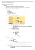
o Overall structure
▪
• CNS – central nervous system – integration system
o Brain + spinal cord
o Afferent signals enter CNS → processed in spinal cord or brain → efferent
signals leave CNS
• PNS – peripheral nervous system
o Input (afferent) system
▪ Somatic senses
▪ Special senses
▪ Visceral senses
o Output (efferent) system
▪ Somatic
▪ Autonomic
• Sympathetic
• Parasympathetic
o Spinal cord
▪ Cross section of the spinal cord
• Grey matter
o Cell bodies
• White matter
o Myelinated axons
▪ Where axons travel in the spinal cord to the brain or out to the
target cells
▪ Afferent system
• Brings signals from the periphery via the dorsal (posterior) horn
▪ Efferent system
• After processing in the spinal cord or brain – efferent fibres carries signals away via
the ventral route
,Neuronal Structure and Function
• Cells of the nervous system – neurons & glia
o Nerve cells (neurons)
▪ Have thread-like extensions
• Dendrites = extensions from the input end
• Axons = extensions from the output end
▪ Different types
• Motor
• Sensory
• Interneurons
▪ Cell bodies are found in grey matter of CNS and in autonomic ganglia
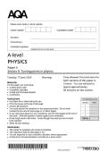
▪ Main structural features
•
o Cell body (soma)
▪ Metabolic engine for the neuron
o Dendrites
▪ Extensions from the input end
o Dendritic spines
▪ Protusions from dendrites that receive input rom a single axon at
the synapse
o Axon hillock
▪ Important trigger zone for action potential
o Axon
▪ Extensions from the output end
o Axon terminals
▪ Synaptic knobs
▪ Makes synaptic connections with other neurons
▪ Types – according to number of processes extending from the cell body
• Multipolar neurons – many processes
• Bipolar neurons – 2 processes
• Unipolar neurons – 1 process
o Pseudo-unipolar neurons
▪ 1 process – typical of sensory neuron
o Glial cells (neuroglia)
▪ Support cells of the CNS
▪ 4 main types
• Astroglia
o Attach to blood vessels
and neurons
o Provide nutrients to
neurons
• Oligodendrocytes
,Neuronal Structure and Function
o Myelin cells
• Microglia
o Important for protecting neurons from infection
• Ependymal cells
o Types
▪ Schwann cells – in PNS
• Myelin cells
▪ Macrophages
o Line fluid filled spaces – ventricles of the brain
▪ Myelin cells
• Schwann cell / oligodendrocyte – myelin sheath
o Schwann cells in PNS
▪ Only covers a single axon
o Oligodendrocytes in CNS
▪ Form myelin around neurons over several axons
• Myelin wraps around axon → forming sheath
• Nerve trunk structure
o Axons
▪ Convey information from neuron to neuron
▪ Organised in bundles – fascicles
▪ Anchored in the connective tissue by the
epineurium
▪ Each bundle is covered by perineurium
▪ Endoneurium – covers individual nerve fibres
• Summary
o Neurons are supported by glia cells
▪ Function of neurons is to transmit information to other neurons or to the neuromuscular
junction via its axon
▪ Neurons are excitable cells – separation of electrical charge across the membrane
• Capable of producing large rapid electrical signals = action potentials
o In PNS – axons are surrounded by schwann cells – which run in peripheral nerve trunks alongside
blood vessels
o Nerves (bundles of axons) can be sensory, motor or mixed
o Peripheral nerves can also contain sympathetic postganglionic fibres – which innervate muscles and
glands
o Power of a neuron depends on its connections
▪ Fast signal transmission – enabled by action potentials
Action Potential
• Objectives
o Describe action potential in terms of:
▪ Ionic basis
▪ Threshold and refractory period
▪ Propagation
• Action potential
o Signal transmission in neurons – structure = function
▪ Axon – has many voltage gated ion channels
▪ Dendrites – has many ligand gated ion channels
o Axon
▪ Electrical transmission down axons
▪ Chemical transmission at synapse – when neuron reaches target cell
, Neuronal Structure and Function
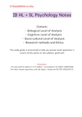
o Action potential
▪ Phases of an action potential
• Resting potential
o Negative membrane
potential
• Depolarisation
o Membrane potential is less
negative than resting
potential
• Repolarisation
o Membrane potential returns from positive resting potential to resting
potential
• Hyperpolarisation
o Membrane potential is more negative than resting potential
▪ Neurons require minimum depolarisation before generating action potential = threshold
▪ Changes in ionic permeability underlie changes in membrane potential
▪ Each action potential is similar in amplitude and duration = all-or-none
▪ Refractory period
• After an action potential has passed along an axon – it cannot conduct another for a
certain period of time
▪ Frequency of action potentials code for stimulus strength
o Fast and slow spiking neurons
▪ Purkinje neuron
• In the cerebellum
• Very short duration = rapid spiking
▪ Pyramidal neuron
• Broader width action potential
• Longer duration spiking
▪ Dopamine neuron
• Long duration
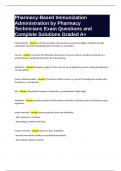
o Threshold for activation
▪ Generation of an action potential
• Requires an adequate stimulus
o Minimum strength required = threshold to open
enough ion channels to bring the membrane
potential to threshold
• Subthreshold = below threshold → no action potential
• Suprathreshold – above threshold → generates action
potential
o Magnitude of the action potential is the same as the
threshold stimulus = all-or-none response
o Rate coding determines stimulus intensity
▪ Strength of stimulus is coded by frequency of action potential
• Ionic basis of the action potential
o Action potentials are fast + unidirectional + simple + effective
o Amplitude is sodium dependent
▪ Less Na+ in extracellular environment = less depolarisation
• Action potential is smaller in amplitude + longer in duration
Structure of the Nervous System
• Objectives
o Understand and name anatomical divisions of the nervous system
o Name and state the function of the main cells of the nervous system – neurons and glia
o Describe the structure of neurons and projections
• Basic anatomical organisation of the nervous system
o Divisions
▪ Brain
▪ Spinal cord
▪ Peripheral nerves
▪ Autonomic nervous system
▪ Enteric nervous system
• Controls activity of the gut
o Overall structure
▪
• CNS – central nervous system – integration system
o Brain + spinal cord
o Afferent signals enter CNS → processed in spinal cord or brain → efferent
signals leave CNS
• PNS – peripheral nervous system
o Input (afferent) system
▪ Somatic senses
▪ Special senses
▪ Visceral senses
o Output (efferent) system
▪ Somatic
▪ Autonomic
• Sympathetic
• Parasympathetic
o Spinal cord
▪ Cross section of the spinal cord
• Grey matter
o Cell bodies
• White matter
o Myelinated axons
▪ Where axons travel in the spinal cord to the brain or out to the
target cells
▪ Afferent system
• Brings signals from the periphery via the dorsal (posterior) horn
▪ Efferent system
• After processing in the spinal cord or brain – efferent fibres carries signals away via
the ventral route
,Neuronal Structure and Function
• Cells of the nervous system – neurons & glia
o Nerve cells (neurons)
▪ Have thread-like extensions
• Dendrites = extensions from the input end
• Axons = extensions from the output end
▪ Different types
• Motor
• Sensory
• Interneurons
▪ Cell bodies are found in grey matter of CNS and in autonomic ganglia
▪ Main structural features
•
o Cell body (soma)
▪ Metabolic engine for the neuron
o Dendrites
▪ Extensions from the input end
o Dendritic spines
▪ Protusions from dendrites that receive input rom a single axon at
the synapse
o Axon hillock
▪ Important trigger zone for action potential
o Axon
▪ Extensions from the output end
o Axon terminals
▪ Synaptic knobs
▪ Makes synaptic connections with other neurons
▪ Types – according to number of processes extending from the cell body
• Multipolar neurons – many processes
• Bipolar neurons – 2 processes
• Unipolar neurons – 1 process
o Pseudo-unipolar neurons
▪ 1 process – typical of sensory neuron
o Glial cells (neuroglia)
▪ Support cells of the CNS
▪ 4 main types
• Astroglia
o Attach to blood vessels
and neurons
o Provide nutrients to
neurons
• Oligodendrocytes
,Neuronal Structure and Function
o Myelin cells
• Microglia
o Important for protecting neurons from infection
• Ependymal cells
o Types
▪ Schwann cells – in PNS
• Myelin cells
▪ Macrophages
o Line fluid filled spaces – ventricles of the brain
▪ Myelin cells
• Schwann cell / oligodendrocyte – myelin sheath
o Schwann cells in PNS
▪ Only covers a single axon
o Oligodendrocytes in CNS
▪ Form myelin around neurons over several axons
• Myelin wraps around axon → forming sheath
• Nerve trunk structure
o Axons
▪ Convey information from neuron to neuron
▪ Organised in bundles – fascicles
▪ Anchored in the connective tissue by the
epineurium
▪ Each bundle is covered by perineurium
▪ Endoneurium – covers individual nerve fibres
• Summary
o Neurons are supported by glia cells
▪ Function of neurons is to transmit information to other neurons or to the neuromuscular
junction via its axon
▪ Neurons are excitable cells – separation of electrical charge across the membrane
• Capable of producing large rapid electrical signals = action potentials
o In PNS – axons are surrounded by schwann cells – which run in peripheral nerve trunks alongside
blood vessels
o Nerves (bundles of axons) can be sensory, motor or mixed
o Peripheral nerves can also contain sympathetic postganglionic fibres – which innervate muscles and
glands
o Power of a neuron depends on its connections
▪ Fast signal transmission – enabled by action potentials
Action Potential
• Objectives
o Describe action potential in terms of:
▪ Ionic basis
▪ Threshold and refractory period
▪ Propagation
• Action potential
o Signal transmission in neurons – structure = function
▪ Axon – has many voltage gated ion channels
▪ Dendrites – has many ligand gated ion channels
o Axon
▪ Electrical transmission down axons
▪ Chemical transmission at synapse – when neuron reaches target cell
, Neuronal Structure and Function
o Action potential
▪ Phases of an action potential
• Resting potential
o Negative membrane
potential
• Depolarisation
o Membrane potential is less
negative than resting
potential
• Repolarisation
o Membrane potential returns from positive resting potential to resting
potential
• Hyperpolarisation
o Membrane potential is more negative than resting potential
▪ Neurons require minimum depolarisation before generating action potential = threshold
▪ Changes in ionic permeability underlie changes in membrane potential
▪ Each action potential is similar in amplitude and duration = all-or-none
▪ Refractory period
• After an action potential has passed along an axon – it cannot conduct another for a
certain period of time
▪ Frequency of action potentials code for stimulus strength
o Fast and slow spiking neurons
▪ Purkinje neuron
• In the cerebellum
• Very short duration = rapid spiking
▪ Pyramidal neuron
• Broader width action potential
• Longer duration spiking
▪ Dopamine neuron
• Long duration
o Threshold for activation
▪ Generation of an action potential
• Requires an adequate stimulus
o Minimum strength required = threshold to open
enough ion channels to bring the membrane
potential to threshold
• Subthreshold = below threshold → no action potential
• Suprathreshold – above threshold → generates action
potential
o Magnitude of the action potential is the same as the
threshold stimulus = all-or-none response
o Rate coding determines stimulus intensity
▪ Strength of stimulus is coded by frequency of action potential
• Ionic basis of the action potential
o Action potentials are fast + unidirectional + simple + effective
o Amplitude is sodium dependent
▪ Less Na+ in extracellular environment = less depolarisation
• Action potential is smaller in amplitude + longer in duration