Introduction to Gastrointestinal Physiology
Structure and Function
• Structure of the GI tract and its associated digestive organs
o Hollow tube starting at the oral cavity and ending at the anal canal
o Each part of the GI tract has a specialised function
▪ Exocrine glands – secretions crucial for digestion and absorption
• Salivary glands
• Liver
• Pancreas
▪ Stomach – storage and breakdown of food
▪ Small intestine – digestion and absorption of food
▪ Large intestine – storage and absorption
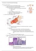
• General structure of the digestive canal
o
▪ Mucosa
• Made up of a layer of epithelial cells – attached to lamina propria – attached to
muscularis mucosae
▪ Submucosa
• Innervated with submucosal plexus – part of enteric nervous system
• Surrounded by muscularis – made up of circular and longitudinal muscles
o Activation of muscles by myenteric plexus = allows activation and
stimulation of muscles – to allow you to mix and propel food down digestive
tract
▪ Serosa
• Connective tissue that holds together muscularis
o Mucosal differences along the GI tract – mucosal layer changes the most as you move across the GI
tract
▪
• Oesophagus
o Layer of tough epithelial cells
o Multiple layer of cells protects oesophagus from potentially damaging food
that is swallowed that moves down from the mouth to the stomach
,Introduction to Gastrointestinal Physiology
• Stomach
o Single layer of columnar epithelial cells dotted with gastric pits
• Small intestine
o Finger like projections (villi) – epithelial surface
o Enable the efficient absorbance of the different components of our diet
• Large intestine
o Flat in nature
o Made up of a single layer of epithelial cells
▪ Involved in absorption of water and electrolytes
o Dotted with goblet cells = mucus secreting cells
▪ Provide mucus to aid in the movement of solid faecal content
o Cell types in the intestinal epithelium
▪ Continuously renewed every three to six days – particularly prone to damage and hypoxia
▪ Originate within the crypts of villi from stem cells
• Stem cells as they move up the crypt become transit amplifying cells = differentiate
into four different cell types → move up through the villi until ejected into the
intestinal lumen
o Absorptive cell
▪ Have microvilli present on the apical surface
o Secretory cells
▪ Goblet cell
• At the apical region of the cytoplasm – full of mucus
secreting granules – when activated = secrete mucus into
intestinal lumen
▪ Enteroendocrine cell
• Packed full of secretory granules on the basal surface of the
cell
o Contain endocrine hormones that are released upon
activation of the cell
▪ Paneth cell
• Packed with secretory granules – lysozyme and antibacterial
agents
o Play a role in defence within the small and large
intestine
• Functions of the GI tract
o Digestion and absorption of nutrients
o Absorption and retention of water and electrolytes
o Elimination of toxins
o Maintenance of barrier function
o Immunological barrier
• Overview of GI function
,Introduction to Gastrointestinal Physiology
o
▪ Oral cavity – secretions
• Salivary glands produce a fluid that is rich in mucus – used in lubrication and
protection of the upper GI tract
• Enzymes required for the initiation of starch digestion
▪ Stomach
• When food is present – stimulates secretions
o HCl = generates low pH in lumen – breaks down contents
o Pepsin enzyme = required for protein digestion
o Intrinsic factor = needed for protecting vitamin B12 for absorption down in
later parts of the small intestine
o Alkaline mucus secretion – for protection of the stomach lumen
• Mixes and breaks up food into the small intestine
▪ Small intestine
• Efficient nutrient digestion and absorption
o Vitamin absorption
o Ion absorption
o Water absorption
▪ Distal small intestine – ileum
• Vitamin B12 absorption
• Bile salt absorption
• Ion and water absorption
▪ Large intestine
• Fine tuning of electrolyte and water absorption
• Daily fluid movements across the GI tract
o
, Introduction to Gastrointestinal Physiology
• Intestinal electrolyte handling: a balance sheet
o
• Splanchnic circulation
o
▪ Represents 20-25% cardiac output
▪ 1.5L/min in fasting state
▪ 3L/min during digestion
▪ Large reservoir – holds 30% blood volume under normal conditions
Control of GI function
• Control of GI function
o 3 different ways
▪ Endocrine control
• Release of hormones from the cells in which they are produced into the circulation
▪ Neurocrine control
• Neurotransmitter release from neurons to act on non-excitatory cells
▪ Paracrine control
• Hormone is released and acts on a local target cell
o Responses
▪ Secretion
▪ Motility
▪ Transport
▪ Release of other hormones
• Gastric acid secretion
o Endocrine, Neurocrine and paracrine mechanisms control gastric acid secretion – control secretion
of HCl
Structure and Function
• Structure of the GI tract and its associated digestive organs
o Hollow tube starting at the oral cavity and ending at the anal canal
o Each part of the GI tract has a specialised function
▪ Exocrine glands – secretions crucial for digestion and absorption
• Salivary glands
• Liver
• Pancreas
▪ Stomach – storage and breakdown of food
▪ Small intestine – digestion and absorption of food
▪ Large intestine – storage and absorption
• General structure of the digestive canal
o
▪ Mucosa
• Made up of a layer of epithelial cells – attached to lamina propria – attached to
muscularis mucosae
▪ Submucosa
• Innervated with submucosal plexus – part of enteric nervous system
• Surrounded by muscularis – made up of circular and longitudinal muscles
o Activation of muscles by myenteric plexus = allows activation and
stimulation of muscles – to allow you to mix and propel food down digestive
tract
▪ Serosa
• Connective tissue that holds together muscularis
o Mucosal differences along the GI tract – mucosal layer changes the most as you move across the GI
tract
▪
• Oesophagus
o Layer of tough epithelial cells
o Multiple layer of cells protects oesophagus from potentially damaging food
that is swallowed that moves down from the mouth to the stomach
,Introduction to Gastrointestinal Physiology
• Stomach
o Single layer of columnar epithelial cells dotted with gastric pits
• Small intestine
o Finger like projections (villi) – epithelial surface
o Enable the efficient absorbance of the different components of our diet
• Large intestine
o Flat in nature
o Made up of a single layer of epithelial cells
▪ Involved in absorption of water and electrolytes
o Dotted with goblet cells = mucus secreting cells
▪ Provide mucus to aid in the movement of solid faecal content
o Cell types in the intestinal epithelium
▪ Continuously renewed every three to six days – particularly prone to damage and hypoxia
▪ Originate within the crypts of villi from stem cells
• Stem cells as they move up the crypt become transit amplifying cells = differentiate
into four different cell types → move up through the villi until ejected into the
intestinal lumen
o Absorptive cell
▪ Have microvilli present on the apical surface
o Secretory cells
▪ Goblet cell
• At the apical region of the cytoplasm – full of mucus
secreting granules – when activated = secrete mucus into
intestinal lumen
▪ Enteroendocrine cell
• Packed full of secretory granules on the basal surface of the
cell
o Contain endocrine hormones that are released upon
activation of the cell
▪ Paneth cell
• Packed with secretory granules – lysozyme and antibacterial
agents
o Play a role in defence within the small and large
intestine
• Functions of the GI tract
o Digestion and absorption of nutrients
o Absorption and retention of water and electrolytes
o Elimination of toxins
o Maintenance of barrier function
o Immunological barrier
• Overview of GI function
,Introduction to Gastrointestinal Physiology
o
▪ Oral cavity – secretions
• Salivary glands produce a fluid that is rich in mucus – used in lubrication and
protection of the upper GI tract
• Enzymes required for the initiation of starch digestion
▪ Stomach
• When food is present – stimulates secretions
o HCl = generates low pH in lumen – breaks down contents
o Pepsin enzyme = required for protein digestion
o Intrinsic factor = needed for protecting vitamin B12 for absorption down in
later parts of the small intestine
o Alkaline mucus secretion – for protection of the stomach lumen
• Mixes and breaks up food into the small intestine
▪ Small intestine
• Efficient nutrient digestion and absorption
o Vitamin absorption
o Ion absorption
o Water absorption
▪ Distal small intestine – ileum
• Vitamin B12 absorption
• Bile salt absorption
• Ion and water absorption
▪ Large intestine
• Fine tuning of electrolyte and water absorption
• Daily fluid movements across the GI tract
o
, Introduction to Gastrointestinal Physiology
• Intestinal electrolyte handling: a balance sheet
o
• Splanchnic circulation
o
▪ Represents 20-25% cardiac output
▪ 1.5L/min in fasting state
▪ 3L/min during digestion
▪ Large reservoir – holds 30% blood volume under normal conditions
Control of GI function
• Control of GI function
o 3 different ways
▪ Endocrine control
• Release of hormones from the cells in which they are produced into the circulation
▪ Neurocrine control
• Neurotransmitter release from neurons to act on non-excitatory cells
▪ Paracrine control
• Hormone is released and acts on a local target cell
o Responses
▪ Secretion
▪ Motility
▪ Transport
▪ Release of other hormones
• Gastric acid secretion
o Endocrine, Neurocrine and paracrine mechanisms control gastric acid secretion – control secretion
of HCl