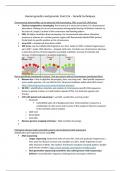
Human genetics and genomic Year3-2a – Genetic techniques.
Chromosomal abnormalities can be detected with karyotyping, FISH, array CGH, SNP arrays
Classical cytogenetics: karyotyping: find numerical or structural variants. For chromosomal
aberrations. Ranking of cut out chromosomes photographed during cell division (mitosis) on
the basis of: Length, Location of the centromere and Banding pattern
FISH: bit higher resolution than karyotyping. For chromosomal aberrations. Detection
presence or absence of a certain genomic region with fluorescently labeled DNA (probe).
Probe binds to specific position on the chromosome
array-CGH = unbalanced aberrations visible
SNP array: can do multiple DNA fragments at a time. Looks for SNPs. Includes fragment just
up to SNP = probe. DNA attaches – elongate with color. Get dots over chromosome: decrease
in intensity and loss of heterozygosity is probably a deletion. Increase in intensity and
increase of heterozygosity is probably duplication.
Not as specifically mentioned in lecture 1 but are ways to look at chromosomes (mentioned later)
Bionano Irys = Way to digitalize karyotyping. Have very long read – label specific sequences
across entire genome. Can use NGS for this. Structural variations visible what NGS cannot.
https://bionanogenomics.com/research/genetic-diseases/
QF-PCR = amplification, detection and analysis of chromosome-specific DNA sequences
known as genetic markers or small tandem repeats (STRs). Too look into zygosity and
trisomy.
CNV with paired end sequencing (= use NGS, usually then use long reads)
o Read-pair
CoNVaDING: gets rid of background noise. CNV prediction is based on a
combination of ratio scores and Z-scores of the sample of interest compared
to the selected control samples.
o Split-read
o Read-depth
o Assembly
Bionano genome mapping technique = high resolution karyotype
Monogenic disease single nucleotide variants can be detected with sequencing
Amplification per fragment (exon) using PCR
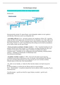
DNA sequencing:
o sanger sequencing: Determine order of every NTs. Only one particular fragment at a
time, gene for disease is known one mutation at a time. Order fragments on size
after inclusion of ddnts. Not random, find known mutation! Get peak pattern. Double
peak means variation. https://www.youtube.com/watch?v=FvHRio1yyhQ
o Next generation sequencing (sometimes also called genome wide sequencing I
believe): no preselection! Sequence whole genome and map to reference.
Chromosomal abnormalities can be detected with karyotyping, FISH, array CGH, SNP arrays
Classical cytogenetics: karyotyping: find numerical or structural variants. For chromosomal
aberrations. Ranking of cut out chromosomes photographed during cell division (mitosis) on
the basis of: Length, Location of the centromere and Banding pattern
FISH: bit higher resolution than karyotyping. For chromosomal aberrations. Detection
presence or absence of a certain genomic region with fluorescently labeled DNA (probe).
Probe binds to specific position on the chromosome
array-CGH = unbalanced aberrations visible
SNP array: can do multiple DNA fragments at a time. Looks for SNPs. Includes fragment just
up to SNP = probe. DNA attaches – elongate with color. Get dots over chromosome: decrease
in intensity and loss of heterozygosity is probably a deletion. Increase in intensity and
increase of heterozygosity is probably duplication.
Not as specifically mentioned in lecture 1 but are ways to look at chromosomes (mentioned later)
Bionano Irys = Way to digitalize karyotyping. Have very long read – label specific sequences
across entire genome. Can use NGS for this. Structural variations visible what NGS cannot.
https://bionanogenomics.com/research/genetic-diseases/
QF-PCR = amplification, detection and analysis of chromosome-specific DNA sequences
known as genetic markers or small tandem repeats (STRs). Too look into zygosity and
trisomy.
CNV with paired end sequencing (= use NGS, usually then use long reads)
o Read-pair
CoNVaDING: gets rid of background noise. CNV prediction is based on a
combination of ratio scores and Z-scores of the sample of interest compared
to the selected control samples.
o Split-read
o Read-depth
o Assembly
Bionano genome mapping technique = high resolution karyotype
Monogenic disease single nucleotide variants can be detected with sequencing
Amplification per fragment (exon) using PCR
DNA sequencing:
o sanger sequencing: Determine order of every NTs. Only one particular fragment at a
time, gene for disease is known one mutation at a time. Order fragments on size
after inclusion of ddnts. Not random, find known mutation! Get peak pattern. Double
peak means variation. https://www.youtube.com/watch?v=FvHRio1yyhQ
o Next generation sequencing (sometimes also called genome wide sequencing I
believe): no preselection! Sequence whole genome and map to reference.