Neural Networks & Reorganisation
LES 2 - Neuroscientific Methods (26/10)
Neuroscientific methods intro
Neuroscientific methods = meting van hersenactiviteit op
systeem niveau, dus niet invasief maar thv de schedel.
Besproken zijn de nodige equipment, neurofysiologische
basis en voorbeelden van toepassingen.
Log size = aandeel van de hersenen dat wordt gemeten
(dus op welk niveau)
MRI grote spacial resolution maar slechte temporal
resolution, EEG omgekeerd.
Magnetic Resonance Imaging (MRI)
What isn’t MRI?
- Not bumpology
Claims that bumps on the skull reflected exaggerated functions/traits
It lacked any mechanism underlying its claims.
It used anecdotal, rather than scientific, evidence.
Nevertheless, its central idea persisted: Localization of Function
- Not mind-reading : MRI kan geen gedachten lezen
- Not invasive: niet intra-craniaal <-> in tegenstelling tot Positron Emission Tomography die wel invasief is
What is MRI?
- Magnetic Resonance Imaging, ofwel MRI, bestaat uit een grote
magneet en head coil
- Drie modaliteiten
o MRI = hersen anatomie
o fMRI = hersenactiviteit
o fc MRI = functionele connectiviteit -> hoe regio’s
geconnecteerd zijn
- Geschiedenis niet op EX
Biologische basis MRI
Meet structureel de hersenanatomie
Heeft te maken met de magnetische kenmerken van protonen in de nuclei van de atomen. Protonen hebben een
massa, zijn positief geladen en draaien rond -> en omdat ze ronddraaien hebben ze een klein maar meetbaar
magnetisch veld. Deze protonen bevinden zich vnl thv water en vetweefsel
, 1. In everyday life, the protons in our body are in balance, randomly
oriented, but in balance.
2. Inside the MRI scanner, which is one giant magnet, the protons
align to the magnetic field (B0). Either in parallel (same direction) or
anti-parallel (opposite direction)
3. The majority of atoms aligns in parallel, allowing to define the NET
magnetisation of the protons in the direction of B0
- Giant Magnet, always on, causes B0 + Head coil, set on or off with a
computer.
- Also called Radio frequency coil, because it emits a radio frequency
4. Now, this is what happens if you are positioned in the scanner. And
this magnetic field is ALWAYS on. See scanner on next slide
5. Emission of a radio frequency pulse by the head coil, induces a flip
of the NET magnetisation (instead of aligning to the Z-axis, the protons
now align in the X-Y field) -> Proton is in ‘excitation state’ = high
energy state
6. However, protons don’t like being in this ‘high-energy’ excitation
state’, and from the moment the radio frequency pulse is turned off, it
will ‘relax’ to its initial position (i.e., align back to the z-axis of the B0
field).
During this ‘relaxation state’, the protons emit radio frequency
themselves, and this signal is measured. (the head coil, both emits and
measures radio frequencies)
-> Proton emits radio frequency during ‘relaxation state’
- Head coil is transmitter & receiver
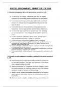
7. The time it takes for a proton to relax to 63% of it’s initial state
(along the z-axis) is called T1. -> snelheid (velocity) is afhankelijk van
het type weefsel
8. Not all tissues ‘relax’ the same way! Protons in fat (e.g. white
matter), relax way faster, than protons in liquid (e.g., cerebrospinal
fluid) By measuring the relaxation in different tissues, contrasts can be
visualized! In so-called ‘T1-weighted’ images, liquid is dark (less energy
emitted), and fat is bright (more energy emitted).
- Darker = lesser energy emitted
Kan in 3D gemeten worden
Toepassingsvoorbeeld : Long Term motor function after neonatal stroke: lesion
localization above all -> clinical: localization of brain lesions/ pre surgical
mapping (vb epilepsie) / prediction of disease progression.
- Vb neonatal stroke + long term motor function -> lesion localization
o Unilateral CP -> more pronounced lesions
,Functional MRI (fMRI)
Biologische basis fMRI
Functional Magnetic Resonance Imaging (fMRI): uses MRI (zelfde
equipment) to indirectly measure brain activity
fMRI is based on the assumption that neuronal activity requires O2
which is carried by the blood; increased blood flow and resulting
hemodynamics are foundation to fMRI.
Achtergrond informatie: localizing brain activity by measuring changes in blood flow is een idee dat al lange tijd
meegaat.
1) Same principle as MRI: contrasts in brain images based on measurement of magnetic relaxation. Here
however, it is not about protons, but about hemoglobin in the blood.
2) Brain region active => increased O2 metabolism => increased blood flow -> fMRI measures the Blood Oxygen
Level Dependent (BOLD) signal
3) Oxyhemoglobin -> diamagnetic (same as tissue)
Deoxyhemoglobin -> paramagnetic (= weak magnetic) interacts with the magnetic signal of the MRI scanner.
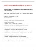
4) BOLD signal
Cel eerst niet geactiveerd door stimulus Toegenomen bloedflow naar die regio iets later
Bij toegenomen stimulatie gaat er meer glucose en O2 Minder gedeoxygeneerd bloed waardoor hoger BOLD
naar het neuron want is meer nodig signaal
Meer gedeoxygeneerd bloed maw lagere BOLD signal
Toegenomen stimulatie occipitaal door visuele prikkel
Initiele dip = toegenomen glucose en O2 consumptie
Gevolgd door:
Delay in respons om bloedflow te laten toenemen (+- 5
seconden)
Kleine overshoot
Increased hemodynamic response
, fMRI always measures a change in BOLD response => you always need a
baseline condition. Most simple design: Block design -> Experimental
condition: stimulus on vs Baseline condition: stimulus off
Heat map activation: Geel = meeste activatie (occipitaal meeste want
visuele stimulus)
Conclusie: deze regio toont meer hersenactiviteit tijdens visuele stimulatie
dan tijdens de baseline -> logisch want is de visuele cortex.
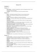
Voorbeeldstudie : Underconnectivity of the superior temporal sulcus predicts emotion recognition deficits in autism
Achtergrondinformatie: studie :
- Actie-perceptie system -> mirror systeem (observatie en execution/ nadoen)
- MNS = motor neuron system interageert met visuele cortex
- Mirror system functions: Action recognition, Action understanding, Action imitation, Empathy
o -> Link with Autism: Difficulties with interpreting other’s behavior/actions + Difficulties with action
imitation
- Gaan ASS ptn minder hun mirror systeem activeren dan controle groep wanner ze biologische bewegingen zien
Basis netwerk bij beiden groepen geactiveerd
In controle groep is de activatie iets meer breder verspreidt
Group comparison Control > ASD -> Significant differences in bilateral superior
temporal sulcus = main visual input region of the human mirror neuron system
Conclusion: Core fronto-parietal regions of human mirror system is largely
intact in ASD, albeit activity in visual input region (STS) is reduced.
Toepassingsvoorbeeld:
- Measurement of fMRI BOLD response (contrast movement > baseline)
- Comparison of contrast values between groups with or without freezing of gait
- Freezers show less recruitment of motor and frontal areas (Studie: The neural correlates of upper limb motor
blocks in Parkinson’s disease and their relation to freezing of gait)
FMRI zoekt naar verschillende hersenregio’s die tot een functioneel netwerk behoren
Sidenote: Discovery of the default mode network... In typical fMRI experiments, researchers are most often interested
in what happens during the experimental condition. But what happens during those ‘rest’ blocks? Is the brain really at
rest?
Default mode network komt online wanneer we in rust zijn -> consistente bevinding doorheen fMRI (frontal,
central, lateral parietal, .. regios)
Probably one of the most consistent findings among fMRI studies was the observation that during ‘rest’ a particular
circuit of brain regions tends to activate and that from the moment a person engages in a particular task, that activity
in these regions decreases.
The network was called the ‘task-negative network’ or ‘default mode network’. Later research showed that activity in
this network remained high when persons are instructed to ‘think of thereselves in relation to others’, ‘mind-wonder’,
‘introspection’...
LES 2 - Neuroscientific Methods (26/10)
Neuroscientific methods intro
Neuroscientific methods = meting van hersenactiviteit op
systeem niveau, dus niet invasief maar thv de schedel.
Besproken zijn de nodige equipment, neurofysiologische
basis en voorbeelden van toepassingen.
Log size = aandeel van de hersenen dat wordt gemeten
(dus op welk niveau)
MRI grote spacial resolution maar slechte temporal
resolution, EEG omgekeerd.
Magnetic Resonance Imaging (MRI)
What isn’t MRI?
- Not bumpology
Claims that bumps on the skull reflected exaggerated functions/traits
It lacked any mechanism underlying its claims.
It used anecdotal, rather than scientific, evidence.
Nevertheless, its central idea persisted: Localization of Function
- Not mind-reading : MRI kan geen gedachten lezen
- Not invasive: niet intra-craniaal <-> in tegenstelling tot Positron Emission Tomography die wel invasief is
What is MRI?
- Magnetic Resonance Imaging, ofwel MRI, bestaat uit een grote
magneet en head coil
- Drie modaliteiten
o MRI = hersen anatomie
o fMRI = hersenactiviteit
o fc MRI = functionele connectiviteit -> hoe regio’s
geconnecteerd zijn
- Geschiedenis niet op EX
Biologische basis MRI
Meet structureel de hersenanatomie
Heeft te maken met de magnetische kenmerken van protonen in de nuclei van de atomen. Protonen hebben een
massa, zijn positief geladen en draaien rond -> en omdat ze ronddraaien hebben ze een klein maar meetbaar
magnetisch veld. Deze protonen bevinden zich vnl thv water en vetweefsel
, 1. In everyday life, the protons in our body are in balance, randomly
oriented, but in balance.
2. Inside the MRI scanner, which is one giant magnet, the protons
align to the magnetic field (B0). Either in parallel (same direction) or
anti-parallel (opposite direction)
3. The majority of atoms aligns in parallel, allowing to define the NET
magnetisation of the protons in the direction of B0
- Giant Magnet, always on, causes B0 + Head coil, set on or off with a
computer.
- Also called Radio frequency coil, because it emits a radio frequency
4. Now, this is what happens if you are positioned in the scanner. And
this magnetic field is ALWAYS on. See scanner on next slide
5. Emission of a radio frequency pulse by the head coil, induces a flip
of the NET magnetisation (instead of aligning to the Z-axis, the protons
now align in the X-Y field) -> Proton is in ‘excitation state’ = high
energy state
6. However, protons don’t like being in this ‘high-energy’ excitation
state’, and from the moment the radio frequency pulse is turned off, it
will ‘relax’ to its initial position (i.e., align back to the z-axis of the B0
field).
During this ‘relaxation state’, the protons emit radio frequency
themselves, and this signal is measured. (the head coil, both emits and
measures radio frequencies)
-> Proton emits radio frequency during ‘relaxation state’
- Head coil is transmitter & receiver
7. The time it takes for a proton to relax to 63% of it’s initial state
(along the z-axis) is called T1. -> snelheid (velocity) is afhankelijk van
het type weefsel
8. Not all tissues ‘relax’ the same way! Protons in fat (e.g. white
matter), relax way faster, than protons in liquid (e.g., cerebrospinal
fluid) By measuring the relaxation in different tissues, contrasts can be
visualized! In so-called ‘T1-weighted’ images, liquid is dark (less energy
emitted), and fat is bright (more energy emitted).
- Darker = lesser energy emitted
Kan in 3D gemeten worden
Toepassingsvoorbeeld : Long Term motor function after neonatal stroke: lesion
localization above all -> clinical: localization of brain lesions/ pre surgical
mapping (vb epilepsie) / prediction of disease progression.
- Vb neonatal stroke + long term motor function -> lesion localization
o Unilateral CP -> more pronounced lesions
,Functional MRI (fMRI)
Biologische basis fMRI
Functional Magnetic Resonance Imaging (fMRI): uses MRI (zelfde
equipment) to indirectly measure brain activity
fMRI is based on the assumption that neuronal activity requires O2
which is carried by the blood; increased blood flow and resulting
hemodynamics are foundation to fMRI.
Achtergrond informatie: localizing brain activity by measuring changes in blood flow is een idee dat al lange tijd
meegaat.
1) Same principle as MRI: contrasts in brain images based on measurement of magnetic relaxation. Here
however, it is not about protons, but about hemoglobin in the blood.
2) Brain region active => increased O2 metabolism => increased blood flow -> fMRI measures the Blood Oxygen
Level Dependent (BOLD) signal
3) Oxyhemoglobin -> diamagnetic (same as tissue)
Deoxyhemoglobin -> paramagnetic (= weak magnetic) interacts with the magnetic signal of the MRI scanner.
4) BOLD signal
Cel eerst niet geactiveerd door stimulus Toegenomen bloedflow naar die regio iets later
Bij toegenomen stimulatie gaat er meer glucose en O2 Minder gedeoxygeneerd bloed waardoor hoger BOLD
naar het neuron want is meer nodig signaal
Meer gedeoxygeneerd bloed maw lagere BOLD signal
Toegenomen stimulatie occipitaal door visuele prikkel
Initiele dip = toegenomen glucose en O2 consumptie
Gevolgd door:
Delay in respons om bloedflow te laten toenemen (+- 5
seconden)
Kleine overshoot
Increased hemodynamic response
, fMRI always measures a change in BOLD response => you always need a
baseline condition. Most simple design: Block design -> Experimental
condition: stimulus on vs Baseline condition: stimulus off
Heat map activation: Geel = meeste activatie (occipitaal meeste want
visuele stimulus)
Conclusie: deze regio toont meer hersenactiviteit tijdens visuele stimulatie
dan tijdens de baseline -> logisch want is de visuele cortex.
Voorbeeldstudie : Underconnectivity of the superior temporal sulcus predicts emotion recognition deficits in autism
Achtergrondinformatie: studie :
- Actie-perceptie system -> mirror systeem (observatie en execution/ nadoen)
- MNS = motor neuron system interageert met visuele cortex
- Mirror system functions: Action recognition, Action understanding, Action imitation, Empathy
o -> Link with Autism: Difficulties with interpreting other’s behavior/actions + Difficulties with action
imitation
- Gaan ASS ptn minder hun mirror systeem activeren dan controle groep wanner ze biologische bewegingen zien
Basis netwerk bij beiden groepen geactiveerd
In controle groep is de activatie iets meer breder verspreidt
Group comparison Control > ASD -> Significant differences in bilateral superior
temporal sulcus = main visual input region of the human mirror neuron system
Conclusion: Core fronto-parietal regions of human mirror system is largely
intact in ASD, albeit activity in visual input region (STS) is reduced.
Toepassingsvoorbeeld:
- Measurement of fMRI BOLD response (contrast movement > baseline)
- Comparison of contrast values between groups with or without freezing of gait
- Freezers show less recruitment of motor and frontal areas (Studie: The neural correlates of upper limb motor
blocks in Parkinson’s disease and their relation to freezing of gait)
FMRI zoekt naar verschillende hersenregio’s die tot een functioneel netwerk behoren
Sidenote: Discovery of the default mode network... In typical fMRI experiments, researchers are most often interested
in what happens during the experimental condition. But what happens during those ‘rest’ blocks? Is the brain really at
rest?
Default mode network komt online wanneer we in rust zijn -> consistente bevinding doorheen fMRI (frontal,
central, lateral parietal, .. regios)
Probably one of the most consistent findings among fMRI studies was the observation that during ‘rest’ a particular
circuit of brain regions tends to activate and that from the moment a person engages in a particular task, that activity
in these regions decreases.
The network was called the ‘task-negative network’ or ‘default mode network’. Later research showed that activity in
this network remained high when persons are instructed to ‘think of thereselves in relation to others’, ‘mind-wonder’,
‘introspection’...