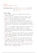
Dorsal-Ventral Axis Formation
Most genes involved in shaping the larval and adult forms of Drosophila identified
by “forward genetics” in the early 1990s
- Randomly mutagenize flies and screen for mutations that disrupted the normal
formation of body plan
- Genes involved in mutant phenotypes were cloned and characterised with
respect to expression patterns and their functions
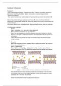
Primary Axis Formation during Oogenesis
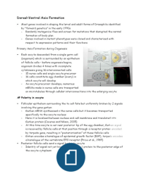
Each oocyte descended from a single germ cell
(oogonium) which is surrounded by an epithelium
of follicle cells – before oogenesis begins,
oogonium divides 4 times with incomplete
cytokinesis giving 16 interconnected cells
- 15 nurse cells and single oocyte precursor
- 16 cells constitute egg chamber (ovary) in
which oocyte will develop
- As oocyte precursor develops, numerous
mRNAs made in nurse cells are transported
on microtubules through cellular interconnections into the enlarging oocyte
AP Polarity in oocyte
Follicular epithelium surrounding the to cell fate but uniformity broken by 2 signals
involving the gene gurken
- Gurken mRNA synthesised n the nurse cells but it becomes transported
specifically to the oocyte nucleus
- Here it is localised between nucleus and cell membrane and translated into
Gurken protein (Caceres and Nilson, 2005)
- At this time oocyte is ver near posterior tip of the egg chamber, Gurken signal
is received by follicle cells at that position through a receptor protein encoded
by torpedo gene, resulting in “posteriorisation” of these follicle cells
- Girken encodes a homologue of epidermal growth factor (EGF), torpedo encodes
a homologue of the vertebrate EFG receptor (Price et al., 1989)
Posterior follicle cells send a signal back into oocyte
- Identity of signal not yet known, recruits par-1 protein to the posterior edge of
the oocyte cytoplasm
, - Par-1 protein organises microtubules with (-) ends
at anterior, (+) ends at posterior ends (Januschke
et al., 2006)
- Orientation of the microtubules is critical because
different microtubule motor proteins will transport their
mRNA or protein cargoes in different directions
Kinesin transported by microtubules to posterior end of
oocyte is oskar mRNA (Zimyanin et al., 2008)
- Oskar mRNA is not able to be translated until it
reaches posterior cortex at which time it
generates the Oskar protein
- Oskar protein recruits more par-1 protein,
stabilising the microtubule orientation and
allowing more material to be recruited to
posterior pole of the oocyte (Doerflinger et al., 2006)
- Posterior pole will have its own distinctive cytoplasm (= pole plasm) which
contains determinants for producing the abdomen and germ cells
Cytoskeletal rearrangement in oocyte is accompanied by an increase in oocyte
volume (due to transfer of contents from nurse cells)
- Components include bicoid and nanos mRNAs, carried by microtubules to anterior
and posterior ends of the oocyte
DV patterning in the oocyte
As oocyte volume increases, oocyte nucleus moves to
anterior dorsal position
- Gurken message becomes localised in a crescent
between oocyte nucleus and the oocyte cell
membrane
- Protein product forms an anterior-posterior gradient along the
dorsal surface of the oocyte
- Gurken protein diffuses a short distance and only reaches the
follicle cells closest to the oocyte nucleus, signalling those cells
to become more dorsal follicle cells (Montell et al., 1991)
- This establishes the dorsal-ventral polarity in the follicle cell
layer that surrounds growing oocyte
Maternal deficiencies of either gurken or torpedo gene causes
ventralisation of the embryo
- Gurken is only active in the oocyte, torpedo is only active in the
somatic follicle cells: revealed by experiments with germline/somatic chimeras
, - Schupbach (1987) transplanted germ cell
precursors from wild-type embryos into
embryos whose mothers carried
torpedo mutation (and vice versa) –
wild-type eggs produced mutant
ventralised embryos when they developed in a
torpedo mutant mother’s egg chamber, torpedo
mutant eggs were able to produce normal
embryos if they developed in a wild-type ovary –
Torpedo protein is needed in the follicle cells,
not in the egg
Gurken-Torpedo signal that specifies dorsalised
follicle cells initiates a cascade of gene activates the
DV axis
1. Activated Torpedo receptor protein inhibits expression of the pipe gene
2. Pipe protein is therefore made only in ventral follicle cells (Sen et al., 1998)
3. Pipe activates Nudel protein (in some unknown mechanism – involves sulphation)
which is secreted to cell membrane of neighbouring ventral embryonic cells
(Zhang et al., 2009)
4. Several hours later, Nudel initiates activation of 3 serine proteases thatare
secreted into perivitelline fluid: proteases are products of gastrulation defective
(gd), snake (snk), easter (ea) genes
- Molecules are secreted in an inactive form and are subsequently activated by
peptide cleavage
- In complex cascade of events – activated Nudel activates the Gastrlation-
defective protease
- Gd protease cleaves the Snake protein, activating the Snake protease which in
turn cleaves the Easter protein
- This cleavage activates the Easter protease, which then cleaves Spätzle protein
5. Cleavage of 3 proteases must only be limited to the most ventral portion of the
embryo
- Accomplished by secretion of protease inhibitor from follicle cells of the ovary
- Inhibitor of Easter and Snake found throughout the perivitelline space
surrounding the embryo (protein very similar to mammalian protease inhibitors
that limit blood clotting protease cascades)
- Proteolytic cleavage of Easter and Spätzle is strictly limited to area around the
most ventral embryonic cells
6. Cleaved Spätzle protein now binds to its receptor in the oocyte cell membrane
(product of the toll gene)
Most genes involved in shaping the larval and adult forms of Drosophila identified
by “forward genetics” in the early 1990s
- Randomly mutagenize flies and screen for mutations that disrupted the normal
formation of body plan
- Genes involved in mutant phenotypes were cloned and characterised with
respect to expression patterns and their functions
Primary Axis Formation during Oogenesis
Each oocyte descended from a single germ cell
(oogonium) which is surrounded by an epithelium
of follicle cells – before oogenesis begins,
oogonium divides 4 times with incomplete
cytokinesis giving 16 interconnected cells
- 15 nurse cells and single oocyte precursor
- 16 cells constitute egg chamber (ovary) in
which oocyte will develop
- As oocyte precursor develops, numerous
mRNAs made in nurse cells are transported
on microtubules through cellular interconnections into the enlarging oocyte
AP Polarity in oocyte
Follicular epithelium surrounding the to cell fate but uniformity broken by 2 signals
involving the gene gurken
- Gurken mRNA synthesised n the nurse cells but it becomes transported
specifically to the oocyte nucleus
- Here it is localised between nucleus and cell membrane and translated into
Gurken protein (Caceres and Nilson, 2005)
- At this time oocyte is ver near posterior tip of the egg chamber, Gurken signal
is received by follicle cells at that position through a receptor protein encoded
by torpedo gene, resulting in “posteriorisation” of these follicle cells
- Girken encodes a homologue of epidermal growth factor (EGF), torpedo encodes
a homologue of the vertebrate EFG receptor (Price et al., 1989)
Posterior follicle cells send a signal back into oocyte
- Identity of signal not yet known, recruits par-1 protein to the posterior edge of
the oocyte cytoplasm
, - Par-1 protein organises microtubules with (-) ends
at anterior, (+) ends at posterior ends (Januschke
et al., 2006)
- Orientation of the microtubules is critical because
different microtubule motor proteins will transport their
mRNA or protein cargoes in different directions
Kinesin transported by microtubules to posterior end of
oocyte is oskar mRNA (Zimyanin et al., 2008)
- Oskar mRNA is not able to be translated until it
reaches posterior cortex at which time it
generates the Oskar protein
- Oskar protein recruits more par-1 protein,
stabilising the microtubule orientation and
allowing more material to be recruited to
posterior pole of the oocyte (Doerflinger et al., 2006)
- Posterior pole will have its own distinctive cytoplasm (= pole plasm) which
contains determinants for producing the abdomen and germ cells
Cytoskeletal rearrangement in oocyte is accompanied by an increase in oocyte
volume (due to transfer of contents from nurse cells)
- Components include bicoid and nanos mRNAs, carried by microtubules to anterior
and posterior ends of the oocyte
DV patterning in the oocyte
As oocyte volume increases, oocyte nucleus moves to
anterior dorsal position
- Gurken message becomes localised in a crescent
between oocyte nucleus and the oocyte cell
membrane
- Protein product forms an anterior-posterior gradient along the
dorsal surface of the oocyte
- Gurken protein diffuses a short distance and only reaches the
follicle cells closest to the oocyte nucleus, signalling those cells
to become more dorsal follicle cells (Montell et al., 1991)
- This establishes the dorsal-ventral polarity in the follicle cell
layer that surrounds growing oocyte
Maternal deficiencies of either gurken or torpedo gene causes
ventralisation of the embryo
- Gurken is only active in the oocyte, torpedo is only active in the
somatic follicle cells: revealed by experiments with germline/somatic chimeras
, - Schupbach (1987) transplanted germ cell
precursors from wild-type embryos into
embryos whose mothers carried
torpedo mutation (and vice versa) –
wild-type eggs produced mutant
ventralised embryos when they developed in a
torpedo mutant mother’s egg chamber, torpedo
mutant eggs were able to produce normal
embryos if they developed in a wild-type ovary –
Torpedo protein is needed in the follicle cells,
not in the egg
Gurken-Torpedo signal that specifies dorsalised
follicle cells initiates a cascade of gene activates the
DV axis
1. Activated Torpedo receptor protein inhibits expression of the pipe gene
2. Pipe protein is therefore made only in ventral follicle cells (Sen et al., 1998)
3. Pipe activates Nudel protein (in some unknown mechanism – involves sulphation)
which is secreted to cell membrane of neighbouring ventral embryonic cells
(Zhang et al., 2009)
4. Several hours later, Nudel initiates activation of 3 serine proteases thatare
secreted into perivitelline fluid: proteases are products of gastrulation defective
(gd), snake (snk), easter (ea) genes
- Molecules are secreted in an inactive form and are subsequently activated by
peptide cleavage
- In complex cascade of events – activated Nudel activates the Gastrlation-
defective protease
- Gd protease cleaves the Snake protein, activating the Snake protease which in
turn cleaves the Easter protein
- This cleavage activates the Easter protease, which then cleaves Spätzle protein
5. Cleavage of 3 proteases must only be limited to the most ventral portion of the
embryo
- Accomplished by secretion of protease inhibitor from follicle cells of the ovary
- Inhibitor of Easter and Snake found throughout the perivitelline space
surrounding the embryo (protein very similar to mammalian protease inhibitors
that limit blood clotting protease cascades)
- Proteolytic cleavage of Easter and Spätzle is strictly limited to area around the
most ventral embryonic cells
6. Cleaved Spätzle protein now binds to its receptor in the oocyte cell membrane
(product of the toll gene)