INTEGRATIVE NEUROSCIENCE // LECTURE 1 // PART 1 //
CHAPTER 7 // DEVELOPMENT, STRUCTURE AND FUNCTION
OF THE BRAIN
→ Relative and absolute brain development
→ Strong increase of the cerebral cortex due to increasing folding of the gray matter
(convolutions) AND strong increase of the white matter (connections between the different
areas of the cerebral cortex)
→ In humans, not the increase in frontal cortex areas (prefrontal cortex, see below)
→ The bigger the brain is, the relative more white matter is needed to have a functional
brain
→ Corpus callosum = Connection between right and left hemisphere
➢ Evolution of the human brain
→ The size increased over time, but now our brain is shrinking (for already 10.000 years)
→ So the brain becomes more efficient and you need a little bit less of neuronal tissue which
is positive, because the brain needs a lot of energy
➢ Except for the prefrontal cortex (PFC)
→ Main functions of PFC:
- Planning complex cognitive behavior
- Personality expression
- Cognitive decision making
- Moderate social behavior
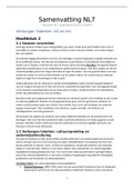
→ Research on human skulls of the past 650 years
a. Present skulls
b. Mid 14th century: Pest victims (black death)
c. 1545: victims of the ship Mary Rose
→ Results:
1. Less pronounced facial features nowadays
2. Increase in cranial vault with 20% from 80 to 95 mm
→ The PFC is getting bigger → You can look at skulls and you can measure the cranial vault
(hersengewelf) and it’s this distance what is found that if you measure that for many skulls,
the present skulls have a lot of skulls from the mid 14th century due to the Pest victims and
,they also found skulls from the ship Mary Rose → If you combine all this, then you find that
you have an increase in the cranial vault with 20% and this is indicative for the increase of
the PFC → Also less pronounced facial features are found in the present skulls
➢ Direction and location indication in 3D
→ Dorsal = back & Ventral = belly
→ Anterior = front & Posterior = back
➢ Anatomical planes of sections and associated terms
→ Frontal or coronal is the same in this plane
➢ Global layout of major brain parts
→ Cerebrum = grote hersenen
→ Cerebellum = kleine hersenen
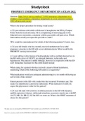
→ In this picture you see the development of the brain
→ The neural plate is depicted in green (most important) from the ectoderm→ Gradually it
changes in shape until it’s called the neural tube which will become the brain and the spinal
cord
→ The neural crest (purple) which is the starting point of the peripheral nervous system
→ So you see the dorsal view of the embryo and there are some other terms like it starts at
the neural plate, then you call it the neural groove and then you see it turns in to the neural
tube
, o ECTODERM
Ectoderm = the outermost layer of cells or tissue of an embryo in early development, or
the parts derived from this
- The surface ectoderm develops into: epidermis, hair, nails, lens of the eye,
sebaceous glands, cornea, tooth enamel, the epithelium of the mouth and nose
- The neural crest of the ectoderm develops into: peripheral nervous system, adrenal
medulla, melanocytes, facial cartilage, dentin of teeth
- The neural tube of the ectoderm develops into: brain, spinal cord, posterior
pituitary, motor neurons, retina
→ Some parts of the brain develop from the ectoderm→ You should know the red terms
→ Ectodermal dysplasia = A growth and developmental disorder of the ectoderm.
→ Many parts of the boy arise from this tissue, such as the skin, sweat glands, teeth and
eyes. The condition is hereditary and occurs in about seven out of then thousand newborns.
The disease occurs in various ways. Anhidrotic ectodermal dysplasia is most common. With
this condition the patients don’t sweat.
◼ MESODERM
Mesoderm = the middle layer of cells or tissue of an embryo in early development, or the
parts derived from this
- The mesoderm forms: muscle (smooth and striated), bone, cartilage, connective
tissue, adipose tissue, circulatory system, lymphatic system, dermis, genitourinary
system, serious membranes, and notochord
◼ ENDODERM
Endoderm = the innermost layer of cells or tissue of an embryo in early development, or
the parts derived from this
- The endoderm forms: the pharynx, the esophagus, the stomach, the small intestine,
the colon, the liver, the pancreas, the bladder, the epithelial parts of the trachea and
bronchi, the lungs, the thyroid and the parathyroid
EXAM QUESTION
From which of the three embryonic germ sheets (endoderm, ectoderm or mesoderm)
develops the peripheral nervous system (PNS) and the central nervous system (CNS)?
a. Ectoderm (CNS and PNS)
b. Endoderm (CNS and PNS)
c. Ectoderm (CNS) and mesoderm (PNS)
d. Ectoderm (CNS) and endoderm (PNS)
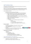
➢ Scanning electron microscopy of a very young embryo
→ Grey picture: You see the folding and the development of the neural tube
→ At the age of 22 – 23 days of the embryo the whole tissue will be closed and things can go
wrong there
, → Developmental disorders of the neural tube.
→ Not closing the neural tube completely leads to Anencephaly (anterior) or Spina bifida
(posterior) (open ruggetje)
→ So the normal situation that it’s totally closed
➢ The next stage: the head of the neural tube differentiates into three parts
→ Forebrain differentiates further into the cerebrum (telencephalon) and midbrain
(diencephalon)
→ Optic vesicles is where the eyes will develop
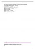
➢ The origin of the retina as a bulge of the diencephalon
→ Early human embryo of approx. 8/9 weeks old
→ Early stage of the cerebrum. The eye is also already under construction
→ Further differentiation of the telencephalon in the left and right half of the brain
(cerebrum). The hindbrain also forms two new parts: the metencephalon (become the
cerebellum) and the spinal cord
➢ Global division of the central nervous system
→ Once the brain is fully developed → You have the global division in to the forebrain,
midbrain and hindbrain
→ Also the spinal cord is divided in to the cervical, thoracic, lumbar and sacral
CHAPTER 7 // DEVELOPMENT, STRUCTURE AND FUNCTION
OF THE BRAIN
→ Relative and absolute brain development
→ Strong increase of the cerebral cortex due to increasing folding of the gray matter
(convolutions) AND strong increase of the white matter (connections between the different
areas of the cerebral cortex)
→ In humans, not the increase in frontal cortex areas (prefrontal cortex, see below)
→ The bigger the brain is, the relative more white matter is needed to have a functional
brain
→ Corpus callosum = Connection between right and left hemisphere
➢ Evolution of the human brain
→ The size increased over time, but now our brain is shrinking (for already 10.000 years)
→ So the brain becomes more efficient and you need a little bit less of neuronal tissue which
is positive, because the brain needs a lot of energy
➢ Except for the prefrontal cortex (PFC)
→ Main functions of PFC:
- Planning complex cognitive behavior
- Personality expression
- Cognitive decision making
- Moderate social behavior
→ Research on human skulls of the past 650 years
a. Present skulls
b. Mid 14th century: Pest victims (black death)
c. 1545: victims of the ship Mary Rose
→ Results:
1. Less pronounced facial features nowadays
2. Increase in cranial vault with 20% from 80 to 95 mm
→ The PFC is getting bigger → You can look at skulls and you can measure the cranial vault
(hersengewelf) and it’s this distance what is found that if you measure that for many skulls,
the present skulls have a lot of skulls from the mid 14th century due to the Pest victims and
,they also found skulls from the ship Mary Rose → If you combine all this, then you find that
you have an increase in the cranial vault with 20% and this is indicative for the increase of
the PFC → Also less pronounced facial features are found in the present skulls
➢ Direction and location indication in 3D
→ Dorsal = back & Ventral = belly
→ Anterior = front & Posterior = back
➢ Anatomical planes of sections and associated terms
→ Frontal or coronal is the same in this plane
➢ Global layout of major brain parts
→ Cerebrum = grote hersenen
→ Cerebellum = kleine hersenen
→ In this picture you see the development of the brain
→ The neural plate is depicted in green (most important) from the ectoderm→ Gradually it
changes in shape until it’s called the neural tube which will become the brain and the spinal
cord
→ The neural crest (purple) which is the starting point of the peripheral nervous system
→ So you see the dorsal view of the embryo and there are some other terms like it starts at
the neural plate, then you call it the neural groove and then you see it turns in to the neural
tube
, o ECTODERM
Ectoderm = the outermost layer of cells or tissue of an embryo in early development, or
the parts derived from this
- The surface ectoderm develops into: epidermis, hair, nails, lens of the eye,
sebaceous glands, cornea, tooth enamel, the epithelium of the mouth and nose
- The neural crest of the ectoderm develops into: peripheral nervous system, adrenal
medulla, melanocytes, facial cartilage, dentin of teeth
- The neural tube of the ectoderm develops into: brain, spinal cord, posterior
pituitary, motor neurons, retina
→ Some parts of the brain develop from the ectoderm→ You should know the red terms
→ Ectodermal dysplasia = A growth and developmental disorder of the ectoderm.
→ Many parts of the boy arise from this tissue, such as the skin, sweat glands, teeth and
eyes. The condition is hereditary and occurs in about seven out of then thousand newborns.
The disease occurs in various ways. Anhidrotic ectodermal dysplasia is most common. With
this condition the patients don’t sweat.
◼ MESODERM
Mesoderm = the middle layer of cells or tissue of an embryo in early development, or the
parts derived from this
- The mesoderm forms: muscle (smooth and striated), bone, cartilage, connective
tissue, adipose tissue, circulatory system, lymphatic system, dermis, genitourinary
system, serious membranes, and notochord
◼ ENDODERM
Endoderm = the innermost layer of cells or tissue of an embryo in early development, or
the parts derived from this
- The endoderm forms: the pharynx, the esophagus, the stomach, the small intestine,
the colon, the liver, the pancreas, the bladder, the epithelial parts of the trachea and
bronchi, the lungs, the thyroid and the parathyroid
EXAM QUESTION
From which of the three embryonic germ sheets (endoderm, ectoderm or mesoderm)
develops the peripheral nervous system (PNS) and the central nervous system (CNS)?
a. Ectoderm (CNS and PNS)
b. Endoderm (CNS and PNS)
c. Ectoderm (CNS) and mesoderm (PNS)
d. Ectoderm (CNS) and endoderm (PNS)
➢ Scanning electron microscopy of a very young embryo
→ Grey picture: You see the folding and the development of the neural tube
→ At the age of 22 – 23 days of the embryo the whole tissue will be closed and things can go
wrong there
, → Developmental disorders of the neural tube.
→ Not closing the neural tube completely leads to Anencephaly (anterior) or Spina bifida
(posterior) (open ruggetje)
→ So the normal situation that it’s totally closed
➢ The next stage: the head of the neural tube differentiates into three parts
→ Forebrain differentiates further into the cerebrum (telencephalon) and midbrain
(diencephalon)
→ Optic vesicles is where the eyes will develop
➢ The origin of the retina as a bulge of the diencephalon
→ Early human embryo of approx. 8/9 weeks old
→ Early stage of the cerebrum. The eye is also already under construction
→ Further differentiation of the telencephalon in the left and right half of the brain
(cerebrum). The hindbrain also forms two new parts: the metencephalon (become the
cerebellum) and the spinal cord
➢ Global division of the central nervous system
→ Once the brain is fully developed → You have the global division in to the forebrain,
midbrain and hindbrain
→ Also the spinal cord is divided in to the cervical, thoracic, lumbar and sacral