BIOD 331 Module 8/Guaranteed A+ Score
Guide
Module 8: The Renal System
8.1: Structure and Function of the Kidney
● Kidneys filter ¼ of average person’s cardiac output. Process essential ions, reabsorb
substances which maintain normal body fluids, excrete waste products through
urine, regulate blood pressure and volume, and stimulate RBC production. Vital for
maintaining homeostasis
● Adult kidney is bean-shaped, about the size of a fist, weighs approx 5 oz. R kidney
lies slightly lower than the L because of the liver’s location just above it. Both
kidneys are mostly protected by the rib cage bc of their locations between the T12
and L3 vertebrae. Kidney’s medial surface is known as the hilus. Hilus is a concave
cleft, where ureters, blood vessels, and nerves enter kidney. Kidney is encapsulated
in an external fibrous capsule and surrounded by fatty connective tissue. Fatty
tissue provides protection from injury & aids to hold the kidney in place. The
kidneys are considered retroperitoneal organs meaning they are situated posterior
to the peritoneal cavity
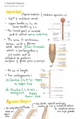
● Nephron is the basic structural & functional unit of a kidney. Approx 1 million
nephrons in each kidney, function to control concentration of water & soluble
materials by filtering the blood, reabsorbing needed materials and excreting waste
products as urine. Nephron eliminates wastes from the body, regulates blood
volume, pH and pressure, and controls the levels of electrolytes. Each nephron
consists of 2 parts, the glomerular capsule (renal corpuscle) and renal tubule. These
structures are connected through the tubule to the associated collecting ducts.
The glomerular capsule filters the blood while the renal tubule reabsorbs needed
materials, and the collecting ducts carry the remaining material away as urine to be
excreted
● ‘]\
● On longitudinal section, the kidney can be divided into the outer cortex and inner
medulla. The outer cortex houses the glomeruli and convoluted tubules (proximal
and distal) of the nephron as well as blood vessels. The inner medulla is comprised
of the Loop of Henle and cone-shaped masses known as the renal pyramids. Portions
of the cortex known as cortical columns project through the medulla to the renal
pyramids. Each pyramid forms a lobe of the kidney. The renal pelvis is located at
, the centermost region of the kidney. It constitutes a funnel-shaped tube that
connects to the ureter as it leaves the hilus. Several extensions of the pelvis called
calyces collect urine which drain continuously into the renal pelvis & subsequently
into the ureter, which transports the urine to the bladder to be stored
● Nephron Structure and Function
○ Functional unit of the kidney, blood filtration & reabsorption take place.
Nephrons can be divided into 1 of 2 groups, cortical nephrons and
juxtamedullary nephrons. Cortical nephrons make up 85% of all nephrons.
They originate superficially in the cortex & have shorter loops of henle that
extend only a short distance into the medulla. Juxtamedullary nephrons
make up remaining 15% of all nephrons. They originate deeper in the cortex,
and their loops of Henle are thinner and extend into the medulla entirely
○ Nephrons receive their blood supply from 2 systems known as the glomerulus
and peritubular capillary network. The glomerulus is a unique system in that
it is located between 2 arterioles, afferent and efferent. Arterioles are
high resistance vessels resulting in an extremely high-pressure system which
can easily force fluid and solutes out of the blood and into the glomerular
capillary along its entire length. The peritubular capillaries are low-pressure
vessels better suited for reabsorption as opposed to filtration. These
capillaries surround the tubules in their entirety allowing rapid movement of
solutes and water. Efferent arterioles located deep in the renal cortex turn
into long, thin-walled vessels known as the vasa recta. The vasa recta run
parallel to the loops of Henle in the medullary region and assist in the
exchange of solutes and water flowing in and out of the kidney
○ The glomerulus is composed of a compact mass of capillaries surrounded by a
thin, double walled capsule known as the Bowman capsule. Blood flows
through the afferent arteriole into the glomerular capillaries, and flows out
of the glomerular capillaries into the efferent arteriole. This then leads to
the peritubular capillaries. Solutes and fluids are filtered from the blood
through the capillary membrane into a fluid-filled space within Bowman
capsule. This space is referred to as Bowman space. The blood that is
filtered into Bowman space is referred to as filtrate. The mass of capillaries
(glomerulus) surrounded by its epithelial capsule (Bowman capsule) that
opens into a tubule is collectively referred to as the renal corpuscle
○ The glomerular capillary membrane contains 3 layers: the capillary endothelial
layer, the basement membrane, and the single-celled capsular epithelial
, layer. The endothelial cells contain small pores called fenestrations which
allow for the filtration of blood. The epithelial layer surrounding the
glomerulus is continuous w the epithelium that lines Bowman capsule.
POdocytes (foot processes) are long extensions of the epithelium that
embed as slit pores, which allow the glomerular filtrate to pass. The
basement membrane is situated between the epithelial and endothelial cell
layers. Spaces within the structural framework of the basement membrane
determine the size-dependent permeability of the glomerulus. The size of these
spaces, under normal circumstances, prevent RBCs and plasma proteins from
passing through the glomerular membrane into the filtrate. A compromise to the
basement membrane would lead to the leakage of RBCs and proteins into the
filtrate which occurs w glomerular disease ○ The nephron tubule is divided into
4 segments
■ The proximal convoluted tubule (highly coiled) which drains Bowman
capsule
■ The loop of Henle
■ The distal convoluted tubule
■ The collecting tubule which joins with other nephron tubules to collect
the filtrate
○ The filtrate will pass through each of these segments before reaching the renal
pelvis. It starts at the proximal convoluted tubule which runs into the descending limb
of the loop of Henle which turns into the ascending loop of Henle as the filtrate returns
to the cortex area. The ascending loop turns into the distal convoluted tubule which
drains into the collecting tubule. The entire tubule is lined w a single layer of epithelial
cells that are situated on a basement membrane. The structure of these cells varies
throughout the tubule allowing for different functions. Epithelial cells in the proximal
tubule are fine and contain villi that increase surface area for reabsorption. They also
contain an increased number of mitochondria which facilitates active transport
processes. In contrast, the epithelial cells of the loop of Henle have fewer mitochondria
leading to less reabsorption and other metabolic processes ● Urine Formation
○ Kidneys filter plasma volume approx 60x each day, using close to 25% of
resting body energy to excrete waste products as urine. Around 47 gallons
of glomerular filtrate containing water, nutrients, and essential ions are
removed daily from the blood plasma. By the time filtrate has entered the
collecting ducts, approx 0.5 gallons of urine has been formed w 99% of
water and nutrients being reabsorbed back into the blood. For this to occur,
Guide
Module 8: The Renal System
8.1: Structure and Function of the Kidney
● Kidneys filter ¼ of average person’s cardiac output. Process essential ions, reabsorb
substances which maintain normal body fluids, excrete waste products through
urine, regulate blood pressure and volume, and stimulate RBC production. Vital for
maintaining homeostasis
● Adult kidney is bean-shaped, about the size of a fist, weighs approx 5 oz. R kidney
lies slightly lower than the L because of the liver’s location just above it. Both
kidneys are mostly protected by the rib cage bc of their locations between the T12
and L3 vertebrae. Kidney’s medial surface is known as the hilus. Hilus is a concave
cleft, where ureters, blood vessels, and nerves enter kidney. Kidney is encapsulated
in an external fibrous capsule and surrounded by fatty connective tissue. Fatty
tissue provides protection from injury & aids to hold the kidney in place. The
kidneys are considered retroperitoneal organs meaning they are situated posterior
to the peritoneal cavity
● Nephron is the basic structural & functional unit of a kidney. Approx 1 million
nephrons in each kidney, function to control concentration of water & soluble
materials by filtering the blood, reabsorbing needed materials and excreting waste
products as urine. Nephron eliminates wastes from the body, regulates blood
volume, pH and pressure, and controls the levels of electrolytes. Each nephron
consists of 2 parts, the glomerular capsule (renal corpuscle) and renal tubule. These
structures are connected through the tubule to the associated collecting ducts.
The glomerular capsule filters the blood while the renal tubule reabsorbs needed
materials, and the collecting ducts carry the remaining material away as urine to be
excreted
● ‘]\
● On longitudinal section, the kidney can be divided into the outer cortex and inner
medulla. The outer cortex houses the glomeruli and convoluted tubules (proximal
and distal) of the nephron as well as blood vessels. The inner medulla is comprised
of the Loop of Henle and cone-shaped masses known as the renal pyramids. Portions
of the cortex known as cortical columns project through the medulla to the renal
pyramids. Each pyramid forms a lobe of the kidney. The renal pelvis is located at
, the centermost region of the kidney. It constitutes a funnel-shaped tube that
connects to the ureter as it leaves the hilus. Several extensions of the pelvis called
calyces collect urine which drain continuously into the renal pelvis & subsequently
into the ureter, which transports the urine to the bladder to be stored
● Nephron Structure and Function
○ Functional unit of the kidney, blood filtration & reabsorption take place.
Nephrons can be divided into 1 of 2 groups, cortical nephrons and
juxtamedullary nephrons. Cortical nephrons make up 85% of all nephrons.
They originate superficially in the cortex & have shorter loops of henle that
extend only a short distance into the medulla. Juxtamedullary nephrons
make up remaining 15% of all nephrons. They originate deeper in the cortex,
and their loops of Henle are thinner and extend into the medulla entirely
○ Nephrons receive their blood supply from 2 systems known as the glomerulus
and peritubular capillary network. The glomerulus is a unique system in that
it is located between 2 arterioles, afferent and efferent. Arterioles are
high resistance vessels resulting in an extremely high-pressure system which
can easily force fluid and solutes out of the blood and into the glomerular
capillary along its entire length. The peritubular capillaries are low-pressure
vessels better suited for reabsorption as opposed to filtration. These
capillaries surround the tubules in their entirety allowing rapid movement of
solutes and water. Efferent arterioles located deep in the renal cortex turn
into long, thin-walled vessels known as the vasa recta. The vasa recta run
parallel to the loops of Henle in the medullary region and assist in the
exchange of solutes and water flowing in and out of the kidney
○ The glomerulus is composed of a compact mass of capillaries surrounded by a
thin, double walled capsule known as the Bowman capsule. Blood flows
through the afferent arteriole into the glomerular capillaries, and flows out
of the glomerular capillaries into the efferent arteriole. This then leads to
the peritubular capillaries. Solutes and fluids are filtered from the blood
through the capillary membrane into a fluid-filled space within Bowman
capsule. This space is referred to as Bowman space. The blood that is
filtered into Bowman space is referred to as filtrate. The mass of capillaries
(glomerulus) surrounded by its epithelial capsule (Bowman capsule) that
opens into a tubule is collectively referred to as the renal corpuscle
○ The glomerular capillary membrane contains 3 layers: the capillary endothelial
layer, the basement membrane, and the single-celled capsular epithelial
, layer. The endothelial cells contain small pores called fenestrations which
allow for the filtration of blood. The epithelial layer surrounding the
glomerulus is continuous w the epithelium that lines Bowman capsule.
POdocytes (foot processes) are long extensions of the epithelium that
embed as slit pores, which allow the glomerular filtrate to pass. The
basement membrane is situated between the epithelial and endothelial cell
layers. Spaces within the structural framework of the basement membrane
determine the size-dependent permeability of the glomerulus. The size of these
spaces, under normal circumstances, prevent RBCs and plasma proteins from
passing through the glomerular membrane into the filtrate. A compromise to the
basement membrane would lead to the leakage of RBCs and proteins into the
filtrate which occurs w glomerular disease ○ The nephron tubule is divided into
4 segments
■ The proximal convoluted tubule (highly coiled) which drains Bowman
capsule
■ The loop of Henle
■ The distal convoluted tubule
■ The collecting tubule which joins with other nephron tubules to collect
the filtrate
○ The filtrate will pass through each of these segments before reaching the renal
pelvis. It starts at the proximal convoluted tubule which runs into the descending limb
of the loop of Henle which turns into the ascending loop of Henle as the filtrate returns
to the cortex area. The ascending loop turns into the distal convoluted tubule which
drains into the collecting tubule. The entire tubule is lined w a single layer of epithelial
cells that are situated on a basement membrane. The structure of these cells varies
throughout the tubule allowing for different functions. Epithelial cells in the proximal
tubule are fine and contain villi that increase surface area for reabsorption. They also
contain an increased number of mitochondria which facilitates active transport
processes. In contrast, the epithelial cells of the loop of Henle have fewer mitochondria
leading to less reabsorption and other metabolic processes ● Urine Formation
○ Kidneys filter plasma volume approx 60x each day, using close to 25% of
resting body energy to excrete waste products as urine. Around 47 gallons
of glomerular filtrate containing water, nutrients, and essential ions are
removed daily from the blood plasma. By the time filtrate has entered the
collecting ducts, approx 0.5 gallons of urine has been formed w 99% of
water and nutrients being reabsorbed back into the blood. For this to occur,