PARTS OF A SPINAL NERVE:
ARISE FROM SC AS ROOTLETS.
Rootlets join to from 2 nerve roots 1. Anterior(ventral) Root, contains motor fibers
2. Posterior(dorsal) Root, contains sensory fibers
Roots unite to form a mixed spinal nerve 1. Motor
2. Sensory
Divides immediately into 2 rami(branches) 1. Anterior(ventral) ramus
2. Posterior(dorsal) ramus
DIAGRAM OF FORMATION OF THE SPINAL NERVE
BRAIN & CRANIAL NERVES:
Contains 3 meningeal layers:
1. DURA MATER- thick external fibrous.
2. ARACHNOID MATER- thin intermediate layer.
3. PIA MATER- delicate internal vaculated layer.
Potential Spaces: Extra dural & Subdural space.
True Spaces: Subarachnoid Space.
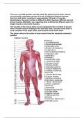
BRAIN DIVIDED INTO 4 MAIN PARTS CEREBRUM, DIENCEPHALON, BRAINSTEM, CEREBELLUM(know positioning
from diag).
BRAINSTEM: Consists of MIDBRAIN, PONS, MEDULLA OBLONGATA.
Functions: Connects cerebrum to SC & cerebellum.
Contains nuclei of majority of cranial nerves & transmits ascending/descending tracts.
CEREBELLUM: Consists of Left & Right Hemispheres.
2nd largest part of brain.
Each hemisphere has narrow parallel
folds cold FOLIA.
Connected to the brainstem.
Functions: Balance, muscle tone,
posture.
DIENCEPHALON: Lies between brainstem &
cerebrum.
Located deep to cerebrum.
Performs vital functions.
3 main parts:
1. Thalamus
2. Hypothalamus
3. Epithalamus
CEREBRUM: Consists of Left & Right
Hemispheres.
ARISE FROM SC AS ROOTLETS.
Rootlets join to from 2 nerve roots 1. Anterior(ventral) Root, contains motor fibers
2. Posterior(dorsal) Root, contains sensory fibers
Roots unite to form a mixed spinal nerve 1. Motor
2. Sensory
Divides immediately into 2 rami(branches) 1. Anterior(ventral) ramus
2. Posterior(dorsal) ramus
DIAGRAM OF FORMATION OF THE SPINAL NERVE
BRAIN & CRANIAL NERVES:
Contains 3 meningeal layers:
1. DURA MATER- thick external fibrous.
2. ARACHNOID MATER- thin intermediate layer.
3. PIA MATER- delicate internal vaculated layer.
Potential Spaces: Extra dural & Subdural space.
True Spaces: Subarachnoid Space.
BRAIN DIVIDED INTO 4 MAIN PARTS CEREBRUM, DIENCEPHALON, BRAINSTEM, CEREBELLUM(know positioning
from diag).
BRAINSTEM: Consists of MIDBRAIN, PONS, MEDULLA OBLONGATA.
Functions: Connects cerebrum to SC & cerebellum.
Contains nuclei of majority of cranial nerves & transmits ascending/descending tracts.
CEREBELLUM: Consists of Left & Right Hemispheres.
2nd largest part of brain.
Each hemisphere has narrow parallel
folds cold FOLIA.
Connected to the brainstem.
Functions: Balance, muscle tone,
posture.
DIENCEPHALON: Lies between brainstem &
cerebrum.
Located deep to cerebrum.
Performs vital functions.
3 main parts:
1. Thalamus
2. Hypothalamus
3. Epithalamus
CEREBRUM: Consists of Left & Right
Hemispheres.