Nutrition and cancer
Lecture 1:
Cancer:
Not one disease
Multistep process
Tumor micro environment
Hallmarks:
Growth and survival:
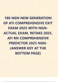
Cell cycle: G1 – S – G2 – M
1. Sustaining proliferative signalling
a. Growth factors (GF) > ex. EGF (gas of car)
b. Up synthesis of GF by tumour cells
c. Up synthesis of GF by neighbouring cells
d. Up receptors at cell surface
e. Structural alterations of receptors ups response
f. Activation of downstream pathway
2. Evading growth suppressors
a. Tumour suppressor genes (break of car)
3. Resisting cell death
a. Necrosis > trigger inflammation
b. Less apoptosis (P53)
4. Enabling replicative immortality
a. Telomere shortening
b. Senescence (viable cell, non-proliferative state, no more
telomeres)
c. Crisis (cell death)
d. Telomerase keeps lengthening telomeres
5. Inducing angiogenesis (new blood vessels)
a. Early stage event
b. Vascular endothelial GF
6. Activating invasion and metastasis (via blood or lymph)
a. Primary tumour formation> local invasion >
intravasation> extravasation> micrometastasis>
macrometastasis
Epithelial to mesenchymal transition: epithelial cells
change to mesenchymal cells (phenotypical change) >
easier to travel > MET when arrived
Enabling characteristics (facilitate other hallmarks)
1. Genome instability and mutations
a. Oncogenes (gas)
b. Tumour suppressor genes (break)
c. Drivers: mutation that drives cancer
2. Tumour promoting inflammation
, a. Supply bioactive molecules
Emerging characteristics
1. Deregulating cellular energetics (aerobic glycolysis> lactate)
2. Avoiding immune destruction
Lecture 2:
Cancer epigenomics: changes in the genome, often affecting gene expression,
not due to changes in DNA sequence (DNA sequence vs structure)
1. DNA methylation
a. Methyl group attached to cystone
b. Sites with C and G (CpG sites) preferred for methylation
c. Transferred to DNA with methyltransferases
d. High frequency of CpG sites is called CpG islands
i. One-carbon metabolism releases methyl group
ii. Methyl groups attached to promotor region > silencing of
gene, preventing binding of transcription factors
iii. Repetitive elements in DNA usually methylated, not
methylated results in unstable genome (hypomethylation)
e. In cancer: silencing of genes resulting from hypermethylation,
relevant in tumour-suppressor genes and DNA repair genes.
f. Methylation can be measured in blood, sputum and urine
2. Histone modifications
a. 4 core histones
b. 1 linker histone
c. Histones have protein tails
d. Groups can bind to tails (phosphate, methyl)
i. Determines how DNA is folded
1. Closed: heterochromatin
2. Open: euchromatin
3. Acetylation: open
4. Methylation: open or closed
3. MiRNA
a. Non-coding RNA
b. Small fragments
c. Regulatory function
d. Aberrant expression in tumours
e. Post-transcriptional regulation of expression
f. Interation with target mRNA
g. Function
i. Translational expression
ii. Cleavage of mRNA
Lecture 3:
Pathology:
Pathologists help diagnose
Take tissue: morphology (eye level), protein level, DNA level
Lecture 1:
Cancer:
Not one disease
Multistep process
Tumor micro environment
Hallmarks:
Growth and survival:
Cell cycle: G1 – S – G2 – M
1. Sustaining proliferative signalling
a. Growth factors (GF) > ex. EGF (gas of car)
b. Up synthesis of GF by tumour cells
c. Up synthesis of GF by neighbouring cells
d. Up receptors at cell surface
e. Structural alterations of receptors ups response
f. Activation of downstream pathway
2. Evading growth suppressors
a. Tumour suppressor genes (break of car)
3. Resisting cell death
a. Necrosis > trigger inflammation
b. Less apoptosis (P53)
4. Enabling replicative immortality
a. Telomere shortening
b. Senescence (viable cell, non-proliferative state, no more
telomeres)
c. Crisis (cell death)
d. Telomerase keeps lengthening telomeres
5. Inducing angiogenesis (new blood vessels)
a. Early stage event
b. Vascular endothelial GF
6. Activating invasion and metastasis (via blood or lymph)
a. Primary tumour formation> local invasion >
intravasation> extravasation> micrometastasis>
macrometastasis
Epithelial to mesenchymal transition: epithelial cells
change to mesenchymal cells (phenotypical change) >
easier to travel > MET when arrived
Enabling characteristics (facilitate other hallmarks)
1. Genome instability and mutations
a. Oncogenes (gas)
b. Tumour suppressor genes (break)
c. Drivers: mutation that drives cancer
2. Tumour promoting inflammation
, a. Supply bioactive molecules
Emerging characteristics
1. Deregulating cellular energetics (aerobic glycolysis> lactate)
2. Avoiding immune destruction
Lecture 2:
Cancer epigenomics: changes in the genome, often affecting gene expression,
not due to changes in DNA sequence (DNA sequence vs structure)
1. DNA methylation
a. Methyl group attached to cystone
b. Sites with C and G (CpG sites) preferred for methylation
c. Transferred to DNA with methyltransferases
d. High frequency of CpG sites is called CpG islands
i. One-carbon metabolism releases methyl group
ii. Methyl groups attached to promotor region > silencing of
gene, preventing binding of transcription factors
iii. Repetitive elements in DNA usually methylated, not
methylated results in unstable genome (hypomethylation)
e. In cancer: silencing of genes resulting from hypermethylation,
relevant in tumour-suppressor genes and DNA repair genes.
f. Methylation can be measured in blood, sputum and urine
2. Histone modifications
a. 4 core histones
b. 1 linker histone
c. Histones have protein tails
d. Groups can bind to tails (phosphate, methyl)
i. Determines how DNA is folded
1. Closed: heterochromatin
2. Open: euchromatin
3. Acetylation: open
4. Methylation: open or closed
3. MiRNA
a. Non-coding RNA
b. Small fragments
c. Regulatory function
d. Aberrant expression in tumours
e. Post-transcriptional regulation of expression
f. Interation with target mRNA
g. Function
i. Translational expression
ii. Cleavage of mRNA
Lecture 3:
Pathology:
Pathologists help diagnose
Take tissue: morphology (eye level), protein level, DNA level