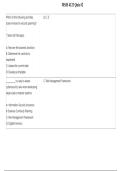
CRNA Study Questions with Correct
Rationale
Venous irritation associated with the injection of
diazepam and lorazepam is secondary to:
1. the high degree of water solubility of these agents
2. the presence of propylene glycol as a solvent
3. the presence of metabisulfite as a preservative
4. the low pH of these agents
Venous irritation associated with the injection of
diazepam and lorazepam is secondary to: the presence of
propylene glycol as a solvent
The insolubility of diazepam and lorazepam in water
requires that parenteral preparations contain propylene
glycol, which has been associated with venous irritation.
In the thromboelastogram below, clot strength is best
represented by:
A
B
E
,F
The maximum amplitude (E) is a measure of the strength
of the fully formed clot. It reflects primarily platelet
number and function although it also requires proper
fibrin formation to achieve normal values.
In patients with a history of hypertrophic
cardiomyopathy, intraoperative management should
include:
1. a nitroglycerine infusion
2. inotropic support
3. afterload reduction
4. maintenance of adequate preload
In patients with a history of hypertrophic
cardiomyopathy, intraoperative management should
include: maintenance of adequate preload
In patients with outflow obstruction, myocardial
depression and maintenance of preload and afterload
are desirable.
Correct statements regarding cerebral metabolism
include:
1. the brain can only utilize glucose as an energy source
, 2. forty percent of brain glucose consumption is
anaerobically metabolized
3. hyperglycemia can reduce the damage from focal
hypoxic injury 4. the adult brain consumes approximately
50 ml/min of oxygen
Correct statements regarding cerebral metabolism
include: the adult brain consumes approximately 50
ml/min of oxygen
The adult brain consumes about 20% of the total body
oxygen (50 ml/min). Neuronal cells normally utilize
glucose as their energy source, but can also utilize ketone
bodies and lactate. Hyperglycemia has been shown to
worsen global and focal hypoxic brain injury.
In the graph of cerebral blood flow below, PaO2 would
best be represented by curve:
1. A
2. B
3. C
4. D
Curve A best represents the effects of changing oxygen
tensions on cerebral blood flow. Hypoxemia causes a