Week 3: Rheumatology
Imaging of inflammatory rheumatic diseases
Arthritis = inflamed joint.
DD painful, swollen joints:
● Gout: (jicht) is an inflammation in the joint (type of arthritis), often in the feet or toe.
○ Erosion can happen in chronic gout → caused by tophi
○ Tophi = deposit of crystalline uric acid at the joint’s surface.
● Pseudo-gout: also inflammation of the joint.
○ It is caused by a deposit of calcium salts in intra-articular cartilage. These
crystals can be seen on ultrasound and X-rays.
○ Often pre-existent osteoarthritis.
Rheumatoid arthritis:
● It can start in one large joint.
● X-rays can show erosion at soon as 3-4 months after diagnosis.
● Conventional X-ray: used to asses erosions and joint space narrowing. Cannot see
synovitis!
● Early RA: Blood test with anti-CCP+, swelling and pain in the joints
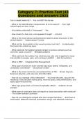
● Difference between osteoarthritis (OA) and RA: OA
= bones grew together in X-ray (no space between
the bones for cartilage) and the bones thicken
(therefore lighter on the X-ray).
● Imaging is needed for early diagnostics, monitoring
efficacy therapy and prognosis.
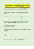
○ Ultrasound: more red on the ultrasound
indicates more Doppler power → more
hyperperfusion (increased blood flow) →
inflammation (synovitis). >>>>>>
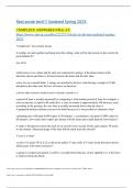
<<<<MRI: white areas on the MRI indicate
water → inflammation. After treatment white
spaces have to become less → monitor the
efficacy of the therapy.
PET: can trace for inflammation or for new bone formation.
● Macrophages: using the folate receptor beta for macrophages.
● B cells: use anti-CD20 (Rituximab)
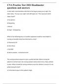
Spondyloarthritis:
● Mainly affect the spine → bamboo spine.
● Inflammation of sacroiliac joint → could eventually
grow together
● New bone formation is used as a tracer for PET
scans.
, Large vessel vasculitis:
● Giant cell arteritis (GCA): inflammation of the lining of the arteries.
● Ultrasound: Halo Sign = wall thickening around the lumen.
Immunology and rheumatic diseases
Problems RA: once a patient has it, it will only get worse. And we do not know how fast it will
get worse.
Development of RA: genetic susceptibility, immune response & trigger.
Diseases:
● Rheumatoid arthritis (RA): Class 2 MHC (HL-DR) and formation of auto-antibodies.
○ Treatment via anti-TNF (Infliximab)
● Ankylosing spondylitis (AS) = axial SpA: Class 1 MHC (HLA-B27) and no formation of
auto-antibodies.
● Henoch-Schonlein purpura: vasculitis of small bloodvesssels. Happens more in
children. Formation of auto-antibodies.
● Systemic lupus erythematosus (SLE): multiple clinical manifestations, like skin, joints,
kidneys etc. Formation of auto-antibodies.
○ Treatment via anti-CD20 (Ritxumab) → works against the lymphoma cells.
○ Or treatment via Type 1 IFN → blocking type 1 IFN (Anifrolumab).
New developments in rheumatology research
The heterogeneity of the diseases hampers diagnosis and therapy, and the need for
research:
● Diagnosis: difficult due to variability and most time after the joint inflammation
● Therapy: many good treatments, but all very expensive. There is still no cure and
there is some non-responsiveness.
Mapping early immune abnormalities in RA:
● There are a lot of environmental factors that play a role before the onset of RA.
● The pre-clinical risk with autoantibodies, serum inflammatory changes and airway
abnormalities.
● Risk factors of the environment are smoking and high BMI.
● Association of arthritis development with synovial T cell infilatrion → indicate
differences in synovium before the onset of disease. But there is no inflammation
seen in this phase → resident T cells.
● These changes could be due to synovial stromal cells, and where are the
auto-antibodies produced in the risk phase?
● In the lymph node in the risk phase, IL-10 goes down → anti-inflammatory.
○ Hypothesis 1: Mesenchymal stromal cells in lymphoid organs and synovial
tissue are defective before the onset of disease → microenvironment with no
good controlled immune response.
○ Hypothesis 2: tolerance lost in other places → production antibodies →
epitope spreading → second hit in joints → chronic arthritis.
Imaging of inflammatory rheumatic diseases
Arthritis = inflamed joint.
DD painful, swollen joints:
● Gout: (jicht) is an inflammation in the joint (type of arthritis), often in the feet or toe.
○ Erosion can happen in chronic gout → caused by tophi
○ Tophi = deposit of crystalline uric acid at the joint’s surface.
● Pseudo-gout: also inflammation of the joint.
○ It is caused by a deposit of calcium salts in intra-articular cartilage. These
crystals can be seen on ultrasound and X-rays.
○ Often pre-existent osteoarthritis.
Rheumatoid arthritis:
● It can start in one large joint.
● X-rays can show erosion at soon as 3-4 months after diagnosis.
● Conventional X-ray: used to asses erosions and joint space narrowing. Cannot see
synovitis!
● Early RA: Blood test with anti-CCP+, swelling and pain in the joints
● Difference between osteoarthritis (OA) and RA: OA
= bones grew together in X-ray (no space between
the bones for cartilage) and the bones thicken
(therefore lighter on the X-ray).
● Imaging is needed for early diagnostics, monitoring
efficacy therapy and prognosis.
○ Ultrasound: more red on the ultrasound
indicates more Doppler power → more
hyperperfusion (increased blood flow) →
inflammation (synovitis). >>>>>>
<<<<MRI: white areas on the MRI indicate
water → inflammation. After treatment white
spaces have to become less → monitor the
efficacy of the therapy.
PET: can trace for inflammation or for new bone formation.
● Macrophages: using the folate receptor beta for macrophages.
● B cells: use anti-CD20 (Rituximab)
Spondyloarthritis:
● Mainly affect the spine → bamboo spine.
● Inflammation of sacroiliac joint → could eventually
grow together
● New bone formation is used as a tracer for PET
scans.
, Large vessel vasculitis:
● Giant cell arteritis (GCA): inflammation of the lining of the arteries.
● Ultrasound: Halo Sign = wall thickening around the lumen.
Immunology and rheumatic diseases
Problems RA: once a patient has it, it will only get worse. And we do not know how fast it will
get worse.
Development of RA: genetic susceptibility, immune response & trigger.
Diseases:
● Rheumatoid arthritis (RA): Class 2 MHC (HL-DR) and formation of auto-antibodies.
○ Treatment via anti-TNF (Infliximab)
● Ankylosing spondylitis (AS) = axial SpA: Class 1 MHC (HLA-B27) and no formation of
auto-antibodies.
● Henoch-Schonlein purpura: vasculitis of small bloodvesssels. Happens more in
children. Formation of auto-antibodies.
● Systemic lupus erythematosus (SLE): multiple clinical manifestations, like skin, joints,
kidneys etc. Formation of auto-antibodies.
○ Treatment via anti-CD20 (Ritxumab) → works against the lymphoma cells.
○ Or treatment via Type 1 IFN → blocking type 1 IFN (Anifrolumab).
New developments in rheumatology research
The heterogeneity of the diseases hampers diagnosis and therapy, and the need for
research:
● Diagnosis: difficult due to variability and most time after the joint inflammation
● Therapy: many good treatments, but all very expensive. There is still no cure and
there is some non-responsiveness.
Mapping early immune abnormalities in RA:
● There are a lot of environmental factors that play a role before the onset of RA.
● The pre-clinical risk with autoantibodies, serum inflammatory changes and airway
abnormalities.
● Risk factors of the environment are smoking and high BMI.
● Association of arthritis development with synovial T cell infilatrion → indicate
differences in synovium before the onset of disease. But there is no inflammation
seen in this phase → resident T cells.
● These changes could be due to synovial stromal cells, and where are the
auto-antibodies produced in the risk phase?
● In the lymph node in the risk phase, IL-10 goes down → anti-inflammatory.
○ Hypothesis 1: Mesenchymal stromal cells in lymphoid organs and synovial
tissue are defective before the onset of disease → microenvironment with no
good controlled immune response.
○ Hypothesis 2: tolerance lost in other places → production antibodies →
epitope spreading → second hit in joints → chronic arthritis.