Unit 3 - Bacteria & Viruses
, Lecture 1 - Bacteria
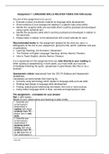
Structure:
↳ protocells are an organised system ofparts with sunstences interacting (in some cases), interior distinct from exterial enviro.
8 can self replicate
↳ Buclesia are prokaryotes:unicellular organisms thatlack nucleir other membrane
enclosed
organelles.
DARYOTIC
CELL: #AMYOTIC
CELL:
e.g. Baclesia, e.g. protists, fungi,
Archea. animals, plants
. non-membune · Have membrane enclosed organelles
enclosed internal
compartments
*ALYOIC
DIVISION:
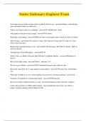
Size of Bacteria:
↳
Binary fission · Antonie van Leeuwenhoek (632-1723) firstobserved
↳ cytokinesis bucleic under microscope.
Imm 110m /nm
Im 10cm 10cm 10 mm 100m
=
=
=
=
= 10 "nm 10A(Angstum)
=
Inm 10 A
=
1 A 0.1nm
=
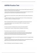
The Gram Stain & the Bacterial Cell Wall:
↳ The Gram stain Method -
separates backlin into groups:NEGATIVE:
aram-positive:violet Dye of layer of
peptidoglycans in periplasmic
3-Differences
-
structure
due to the -
This
-
Gram-negative: Red
Dye the cell wall. space between cell membrane another
·
-
GHAM-POSTTIVE: outer membrane.
-
Thick layer of peptidoglycan outside ofcell Membrane picksup counterstain appeals pink -
red.
-
Thick peptidoglycan cell wall that retains violet dye s
appeals deep blue/purple
, Lecture 1 - Bacteria
Structure:
↳ protocells are an organised system ofparts with sunstences interacting (in some cases), interior distinct from exterial enviro.
8 can self replicate
↳ Buclesia are prokaryotes:unicellular organisms thatlack nucleir other membrane
enclosed
organelles.
DARYOTIC
CELL: #AMYOTIC
CELL:
e.g. Baclesia, e.g. protists, fungi,
Archea. animals, plants
. non-membune · Have membrane enclosed organelles
enclosed internal
compartments
*ALYOIC
DIVISION:
Size of Bacteria:
↳
Binary fission · Antonie van Leeuwenhoek (632-1723) firstobserved
↳ cytokinesis bucleic under microscope.
Imm 110m /nm
Im 10cm 10cm 10 mm 100m
=
=
=
=
= 10 "nm 10A(Angstum)
=
Inm 10 A
=
1 A 0.1nm
=
The Gram Stain & the Bacterial Cell Wall:
↳ The Gram stain Method -
separates backlin into groups:NEGATIVE:
aram-positive:violet Dye of layer of
peptidoglycans in periplasmic
3-Differences
-
structure
due to the -
This
-
Gram-negative: Red
Dye the cell wall. space between cell membrane another
·
-
GHAM-POSTTIVE: outer membrane.
-
Thick layer of peptidoglycan outside ofcell Membrane picksup counterstain appeals pink -
red.
-
Thick peptidoglycan cell wall that retains violet dye s
appeals deep blue/purple