Ribosomes – 70s – smaller than in eukaryotic cells
Less than 2 micrometres
S IMILARITIES TO E UKARYOTIC CELLS
plasma membrane
cytoplasm
ribosomes for protein synthesis
DNA + RNA
DIFFERENCES TO EUKARYOTIC CELLS
no nucleus
no membrane bound organelles
have 70s ribosomes
cell wall made of peptidoglycan – not cellulose/chitin
less well developed cytoplasm – no centrioles
some bacterial cells also have a slime/waxy capsule, plasmids, flagella, pili
BINARY FISSION – BACTERIAL REPRODUCTION
interact by means of a pilus
o thin appendage used to connect the two cells
1) Cell replicates its DNA
2) Cytoplasmic membrane elongates – separates DNA molecules
3) Cross wall forms – membrane invaginates
4) Cross wall forms completely
5) Daughter cells
MICROSCOPES
Microscope – optical instrument used to produce an enlarged image of small
objects
Object – material under microscope
Image – appearance of object viewed through microscope
UNITS OF MEASURE
millimetres (mm)
micrometres (µm)
nanometres (nm)
1 metre – 1000mm
1 mm – 1000 µm
1 µm – 1000nm
MAGNIFICATION
number of times larger an image appears compared to the size of the object
microscopes produce linear magnification
o If the specimen is magnified x100 it's 100 times longer & wider than it
really is
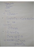
CALCULATING MAGNIFICATION
, I
Magnification=Image ¿ ¿ Actual ¿ ¿ ¿ ¿ M=
A
Nanometres & micrometres – usual units for magnification questions
RESOLUTION
the clarity of the image
the higher the resolution the clearer the image
if the image isn't clear magnification won't help
resolution - the ability of a microscope to distinguish two adjacent points as
separate from one another
maximum resolution - the least distance between 2 closely opposed points at
which they may be recognised as 2 separate entities
the better the resolution – the better you are able to view & identify finer
details
High frequency
o Short wavelength
o Good Resolution
Low frequency
o Long wavelength
o Poor resolution
Resolution dependent on the wavelength of the beam we’re using to see the
material
electron beam is able to provide better detail than light microscopes
o because of its shorter wavelengths
as wavelength gets smaller – ability to resolve the spots gets better
visible light has a longer wavelength than electron beams
LIGHT MICROSCOPE
uses visible light & a system of lenses to magnify images
simplest microscope
cheap, easy to use, portable & can study whole specimens
uses a number of lenses to focus a beam of light
To view the specimens at different magnifications: - light microscopes have
3/4 objective lenses
o x4
o x10
o x40
o x 100 - oil immersion lens
o can be rotated in to position
o eyepiece lens then magnifies the image again - x10
Total magnification = objective lens x eyepiece lens
Max magnification = x1500
Resolution = 200mm (resolution is limited).
Specimens - a wide range of living organisms & sections of organisms can be
viewed
Photomicrograph - photograph of the image seen with a light microscope
Less than 2 micrometres
S IMILARITIES TO E UKARYOTIC CELLS
plasma membrane
cytoplasm
ribosomes for protein synthesis
DNA + RNA
DIFFERENCES TO EUKARYOTIC CELLS
no nucleus
no membrane bound organelles
have 70s ribosomes
cell wall made of peptidoglycan – not cellulose/chitin
less well developed cytoplasm – no centrioles
some bacterial cells also have a slime/waxy capsule, plasmids, flagella, pili
BINARY FISSION – BACTERIAL REPRODUCTION
interact by means of a pilus
o thin appendage used to connect the two cells
1) Cell replicates its DNA
2) Cytoplasmic membrane elongates – separates DNA molecules
3) Cross wall forms – membrane invaginates
4) Cross wall forms completely
5) Daughter cells
MICROSCOPES
Microscope – optical instrument used to produce an enlarged image of small
objects
Object – material under microscope
Image – appearance of object viewed through microscope
UNITS OF MEASURE
millimetres (mm)
micrometres (µm)
nanometres (nm)
1 metre – 1000mm
1 mm – 1000 µm
1 µm – 1000nm
MAGNIFICATION
number of times larger an image appears compared to the size of the object
microscopes produce linear magnification
o If the specimen is magnified x100 it's 100 times longer & wider than it
really is
CALCULATING MAGNIFICATION
, I
Magnification=Image ¿ ¿ Actual ¿ ¿ ¿ ¿ M=
A
Nanometres & micrometres – usual units for magnification questions
RESOLUTION
the clarity of the image
the higher the resolution the clearer the image
if the image isn't clear magnification won't help
resolution - the ability of a microscope to distinguish two adjacent points as
separate from one another
maximum resolution - the least distance between 2 closely opposed points at
which they may be recognised as 2 separate entities
the better the resolution – the better you are able to view & identify finer
details
High frequency
o Short wavelength
o Good Resolution
Low frequency
o Long wavelength
o Poor resolution
Resolution dependent on the wavelength of the beam we’re using to see the
material
electron beam is able to provide better detail than light microscopes
o because of its shorter wavelengths
as wavelength gets smaller – ability to resolve the spots gets better
visible light has a longer wavelength than electron beams
LIGHT MICROSCOPE
uses visible light & a system of lenses to magnify images
simplest microscope
cheap, easy to use, portable & can study whole specimens
uses a number of lenses to focus a beam of light
To view the specimens at different magnifications: - light microscopes have
3/4 objective lenses
o x4
o x10
o x40
o x 100 - oil immersion lens
o can be rotated in to position
o eyepiece lens then magnifies the image again - x10
Total magnification = objective lens x eyepiece lens
Max magnification = x1500
Resolution = 200mm (resolution is limited).
Specimens - a wide range of living organisms & sections of organisms can be
viewed
Photomicrograph - photograph of the image seen with a light microscope