Fractures and Trauma
Medicine
Seán Keenan
2022
,Osteoporosis
Description
Osteoporosis is a reduction in overall bone mass and may be 1o (age related) or 2o to other conditions or drugs. 18 %
of females (80 % of hip fractures are in women >50 YO) and 6 % of males >50 YO are affected. Trabeculae are lost in
women’s bones over time but in men it is mostly conserved.
Osteoporosis
Presentation Management
- Trabecular: Vertebral crush injury; Dowager’s hump - Lifestyle modifications
- Cortical: Long bone fractures; Femoral neck common o ↓Risks: Smoking cessation; Alcohol reduction
- NB: Femoral neck is biggest risk of death o Diet: ↑ Calcium + VitD; ↑ BMI
Causes o Home Aids: ↓ Fall risks
- Important: RA; Corticosteroids; ↑↑ Levothyroxine o Exercises: Weigh bearing; Balance
- Age: Bone mineral density decreases with age - Pharmacological
- Lifestyle: Alcohol >4 units daily; BMI <19 o Key: If Fragility fracture skip DEXA scan to treat
- Systemic: Menopause; CKD; DM o Supplements: Calcium + VitD
- Activity: Prolonged immobility; Falls o Bisphosphonate: Alendronate (CI: GFR<35; PUD)
- Drugs: Antiepileptics; PPIs; Thiazolinediones o Denosumab: RANKLi; Preferred if GFR <30
- NB: SSRIs ↑ risk of # but do not affect bone density o Strontium Ranelate: Last line (CI: CVD)
Investigations o Raloxifene: Selective oestrogen receptor mod.
- Bloods: Ca2+; PO43-; ALP normal o HRT: Post-menopause (CI: Breast Ca; CVD)
- Bone densitometry: Bone mineral density (BMD) o Teriparatide: Recombinant PTH (SE: Renal Ca)
- DEXA: <2.5 SD below young adult mean density o Calcitonin: ↓ Pain after vertebral fracture
- NB: See table below for DEXA scoring - Steroid Therapy
- T-Score: Based on bone mass of young population o Risk: Significant if ≥75 mg for 3 months
- Z-Score: Age; Gender; Ethnic adjusted score o Key: Start bone protection immediately
- X-Ray: Low sensitivity and specificity
- FRAX: 10 yr risk of fragility fracture
- NB: Assess all women ≥65 YO and ≥75 males
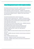
Osteoporosis Risk Factors DEXA Bone Densiometry WHO Criteria
Description T-Score Description
S Steroid use of >5 mg/d of Prednisolone >0 BMD is better than the reference
H Hyperthyroidism; Hyperparathyroidism; Hypercaliuria 0 → -1 BMD is in top 84 % (no evidence of osteoporosis)
A Alcohol + Tobacco use -1 → -2.5 Osteopenia (risk of later osteoporotic fracture)
T Thin (BMI <18.5) < -2.5 Osteoporosis (Repeat DEXA scan every 2 years)
T Testosterone deficit
T-Score vs Z-Score Comparisons
E Early menopause
T-Score: T = Teens (young adults)
R Renal or Liver failure
E Erosive; Inflammatory bone disease
Z-Score: Z = Everything (from A to Z)
D Dietary Calcium Deficiency/Malabsorption; T1DM
, Fractures in General
Description
Bony injury resulting in a fracture may arise from trauma (excessive forces applied to bone), stress related (repetitive
low velocity injury) or pathological (abnormal bone which fractures during normal use of following minimal trauma).
Radiological Description of Fractures
Fracture Site Fracture Description
- Bone: Specific bone fractured - Stable: Bones are still aligned
- Region: Break into ⅓; Proximal; Shaft; Distal - Open: Bone exposed to outside environment
- Joint: Intra-articular involvement - Transverse: Horizontal fracture line
- Epiphyseal: Epiphyseal involvement; Salter-Harris class - Spiral: Fracture line spirals down bone
- Occult #: Not viewable on XR; ∆ on MRI - Oblique: Angled fracture line
Fracture Displacement - Comminuted: Bone shattered into ≥3 pieces
- Degree: Described as % of displacement Paediatric Special Fractures
- Rotation: Rotated from original position - Greenstick: Cortex fractures; Periosteum intact
- Angulated: Fracture angled from breakage - NB: Occur in children due to bone flexibility
- Translation: Bones are overlapping - Spiral: Indicates abuse
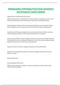
Fracture Types
Fracture Type Description
Oblique Fracture Fracture lies obliquely to long axis of bone
Comminuted Fracture >2 Fragments
Segmental Fracture More than one fracture along a bone
Transverse Fracture Perpendicular to long axis of bone
Spiral Fracture Severe oblique fractures with rotation along long azis of bone
Gustilo and Anderson Classification System
Grade Injury
1 Low energy wound <1 cm
2 Greater than 1 cm wound with moderate soft tissue damage
3 High energy wound >1 cm with extensive soft tissue damage
3A Adequate soft tissue coverage
3B Inadequate soft tissue coverage
3C Associated arterial injury
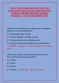
Salter-Harris Classification
Class Description
I Seen in babies or pathological conditions (e.g. scurvy)
II Commonest injury with fracyre line above the growth plate
III There is a displaced fragment with fracture line through the growth plate
IV Union across the growth plate may interfere with bone growth
V Compression of the epiphysis causes deformity and stunting
Medicine
Seán Keenan
2022
,Osteoporosis
Description
Osteoporosis is a reduction in overall bone mass and may be 1o (age related) or 2o to other conditions or drugs. 18 %
of females (80 % of hip fractures are in women >50 YO) and 6 % of males >50 YO are affected. Trabeculae are lost in
women’s bones over time but in men it is mostly conserved.
Osteoporosis
Presentation Management
- Trabecular: Vertebral crush injury; Dowager’s hump - Lifestyle modifications
- Cortical: Long bone fractures; Femoral neck common o ↓Risks: Smoking cessation; Alcohol reduction
- NB: Femoral neck is biggest risk of death o Diet: ↑ Calcium + VitD; ↑ BMI
Causes o Home Aids: ↓ Fall risks
- Important: RA; Corticosteroids; ↑↑ Levothyroxine o Exercises: Weigh bearing; Balance
- Age: Bone mineral density decreases with age - Pharmacological
- Lifestyle: Alcohol >4 units daily; BMI <19 o Key: If Fragility fracture skip DEXA scan to treat
- Systemic: Menopause; CKD; DM o Supplements: Calcium + VitD
- Activity: Prolonged immobility; Falls o Bisphosphonate: Alendronate (CI: GFR<35; PUD)
- Drugs: Antiepileptics; PPIs; Thiazolinediones o Denosumab: RANKLi; Preferred if GFR <30
- NB: SSRIs ↑ risk of # but do not affect bone density o Strontium Ranelate: Last line (CI: CVD)
Investigations o Raloxifene: Selective oestrogen receptor mod.
- Bloods: Ca2+; PO43-; ALP normal o HRT: Post-menopause (CI: Breast Ca; CVD)
- Bone densitometry: Bone mineral density (BMD) o Teriparatide: Recombinant PTH (SE: Renal Ca)
- DEXA: <2.5 SD below young adult mean density o Calcitonin: ↓ Pain after vertebral fracture
- NB: See table below for DEXA scoring - Steroid Therapy
- T-Score: Based on bone mass of young population o Risk: Significant if ≥75 mg for 3 months
- Z-Score: Age; Gender; Ethnic adjusted score o Key: Start bone protection immediately
- X-Ray: Low sensitivity and specificity
- FRAX: 10 yr risk of fragility fracture
- NB: Assess all women ≥65 YO and ≥75 males
Osteoporosis Risk Factors DEXA Bone Densiometry WHO Criteria
Description T-Score Description
S Steroid use of >5 mg/d of Prednisolone >0 BMD is better than the reference
H Hyperthyroidism; Hyperparathyroidism; Hypercaliuria 0 → -1 BMD is in top 84 % (no evidence of osteoporosis)
A Alcohol + Tobacco use -1 → -2.5 Osteopenia (risk of later osteoporotic fracture)
T Thin (BMI <18.5) < -2.5 Osteoporosis (Repeat DEXA scan every 2 years)
T Testosterone deficit
T-Score vs Z-Score Comparisons
E Early menopause
T-Score: T = Teens (young adults)
R Renal or Liver failure
E Erosive; Inflammatory bone disease
Z-Score: Z = Everything (from A to Z)
D Dietary Calcium Deficiency/Malabsorption; T1DM
, Fractures in General
Description
Bony injury resulting in a fracture may arise from trauma (excessive forces applied to bone), stress related (repetitive
low velocity injury) or pathological (abnormal bone which fractures during normal use of following minimal trauma).
Radiological Description of Fractures
Fracture Site Fracture Description
- Bone: Specific bone fractured - Stable: Bones are still aligned
- Region: Break into ⅓; Proximal; Shaft; Distal - Open: Bone exposed to outside environment
- Joint: Intra-articular involvement - Transverse: Horizontal fracture line
- Epiphyseal: Epiphyseal involvement; Salter-Harris class - Spiral: Fracture line spirals down bone
- Occult #: Not viewable on XR; ∆ on MRI - Oblique: Angled fracture line
Fracture Displacement - Comminuted: Bone shattered into ≥3 pieces
- Degree: Described as % of displacement Paediatric Special Fractures
- Rotation: Rotated from original position - Greenstick: Cortex fractures; Periosteum intact
- Angulated: Fracture angled from breakage - NB: Occur in children due to bone flexibility
- Translation: Bones are overlapping - Spiral: Indicates abuse
Fracture Types
Fracture Type Description
Oblique Fracture Fracture lies obliquely to long axis of bone
Comminuted Fracture >2 Fragments
Segmental Fracture More than one fracture along a bone
Transverse Fracture Perpendicular to long axis of bone
Spiral Fracture Severe oblique fractures with rotation along long azis of bone
Gustilo and Anderson Classification System
Grade Injury
1 Low energy wound <1 cm
2 Greater than 1 cm wound with moderate soft tissue damage
3 High energy wound >1 cm with extensive soft tissue damage
3A Adequate soft tissue coverage
3B Inadequate soft tissue coverage
3C Associated arterial injury
Salter-Harris Classification
Class Description
I Seen in babies or pathological conditions (e.g. scurvy)
II Commonest injury with fracyre line above the growth plate
III There is a displaced fragment with fracture line through the growth plate
IV Union across the growth plate may interfere with bone growth
V Compression of the epiphysis causes deformity and stunting