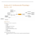
Study unit 3: Cardiovascular Physiology –
SU 3.1 – 3.5
Study Unit 3.1
Cardiovascular System: Components
1. Heart
• Chambers
• Valves
• Conduction and contractile tissue
2. Blood vessels
• Veins, arteries, and capillaries
• Pulmonary, systemic and cardiac circulation
3. Blood
• Cells and plasma
4. Lymphatic system
Cardiovascular System Structure
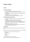
- The cardiovascular system is a closed loop. The heart is a pump that circulates blood through the
system.
,IMPORTANT:
1. Arteries take blood away from the heart
2. Veins carry blood back to the heart.
- Exceptions:
o Hepatic portal system
o Hypophyseal portal system
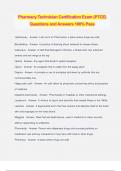
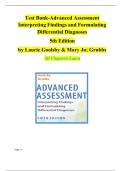
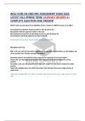
,Anatomy of the thoracic cavity:
Structure of the Heart
- Hollow muscular organ.
- Walls are composed of cardiac muscle (the myocardium).
- Myocardium is lined by an endothelial layer called the endocardium
(in contact with the blood inside the heart cavity)
- Covered by a thin layer called the epicardium (=visceral layer of the
pericardial sac).
- Pericardium: double layered fibroserous sac which covers the heart.
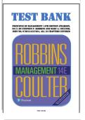
, Contractions of the heart:
The heartbeat happens as follows:
1. The SA node (called the pacemaker of the heart) sends out an electrical impulse.
2. The upper heart chambers (atria) contract.
3. The AV node sends an impulse into the ventricles.
4. The lower heart chambers (ventricles) contract or pump.
5. The SA node sends another signal to the atria to contract, which starts the cycle over again.
Heart Chambers
- The human HEART consists of four chambers:
o Two atria (right and left) which are separated from each other by the interatrial septum.
o Two ventricles (right and left) which are separated from each other by the interventricular
septum. The wall of the left ventricle is about 3 times thicker than the wall of the right
ventricle.
- The ventricular myocardium (wall) is much thicker and stronger than the atrial myocardium (wall).
The atrial muscle (of both atria) is completely separated from the ventricular muscle (of both
ventricles) by a fibrous ring called AV ring (atrioventricular ring).
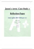
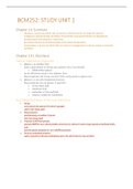
Heart Valves
- Function: Ensure One-Way Flow in the Heart
- Two sets of heart valves ensure one-way flow
1. Atrioventricular valves
• Between atria and ventricles
• Tricuspid valve on the right side
• Bicuspid valve, or mitral valve, on the left side
2. Semilunar valves
• Between ventricles and arteries
• Aortic valve
• Pulmonary valve
SU 3.1 – 3.5
Study Unit 3.1
Cardiovascular System: Components
1. Heart
• Chambers
• Valves
• Conduction and contractile tissue
2. Blood vessels
• Veins, arteries, and capillaries
• Pulmonary, systemic and cardiac circulation
3. Blood
• Cells and plasma
4. Lymphatic system
Cardiovascular System Structure
- The cardiovascular system is a closed loop. The heart is a pump that circulates blood through the
system.
,IMPORTANT:
1. Arteries take blood away from the heart
2. Veins carry blood back to the heart.
- Exceptions:
o Hepatic portal system
o Hypophyseal portal system
,Anatomy of the thoracic cavity:
Structure of the Heart
- Hollow muscular organ.
- Walls are composed of cardiac muscle (the myocardium).
- Myocardium is lined by an endothelial layer called the endocardium
(in contact with the blood inside the heart cavity)
- Covered by a thin layer called the epicardium (=visceral layer of the
pericardial sac).
- Pericardium: double layered fibroserous sac which covers the heart.
, Contractions of the heart:
The heartbeat happens as follows:
1. The SA node (called the pacemaker of the heart) sends out an electrical impulse.
2. The upper heart chambers (atria) contract.
3. The AV node sends an impulse into the ventricles.
4. The lower heart chambers (ventricles) contract or pump.
5. The SA node sends another signal to the atria to contract, which starts the cycle over again.
Heart Chambers
- The human HEART consists of four chambers:
o Two atria (right and left) which are separated from each other by the interatrial septum.
o Two ventricles (right and left) which are separated from each other by the interventricular
septum. The wall of the left ventricle is about 3 times thicker than the wall of the right
ventricle.
- The ventricular myocardium (wall) is much thicker and stronger than the atrial myocardium (wall).
The atrial muscle (of both atria) is completely separated from the ventricular muscle (of both
ventricles) by a fibrous ring called AV ring (atrioventricular ring).
Heart Valves
- Function: Ensure One-Way Flow in the Heart
- Two sets of heart valves ensure one-way flow
1. Atrioventricular valves
• Between atria and ventricles
• Tricuspid valve on the right side
• Bicuspid valve, or mitral valve, on the left side
2. Semilunar valves
• Between ventricles and arteries
• Aortic valve
• Pulmonary valve