Klopto
4: Trauma en gonioscopie
De anatomie en fysiologie van de voorste oogkamer uit te leggen
Afvoer kamerwater:
1. Trabekelsysteem
2. Uveo scleraal
3. Iris
De structuren van de voorste oogkamer, te benoemen en te herkennen
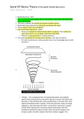
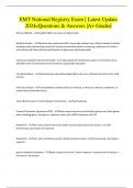
Structuren die we kunnen zien:
- I = iris
- Can’t= ciliary body band (aanhechting iris aan corpus ciliare)
- See = scleraal spoor (witte band)
- Till = trabeculair systeem (ATM en PTM = de afvoer)
- Schwalbers line = overgang tussen cornea en de afvoer. Dus het einde van descemet
Schwalbe’s lijn:
- Kleur varieert van helder (wit/ geel tot lichtbruin) vanwege de pigmentatie
- Maak optic section, verlichtingssysteem 5 tot 10 graden
- Kijk naar 2 reflecties
, 1. Reflex in de kamerhoek
2. Reflex cornea/ sclera
- Schwalbe’s lijn met pigment wordt Sampaolesi lijn
Kanaal van schlemm:
- Circulair veneus kanaal (ligt in sclera net achter/ onder het limbus
gebied)
- Bevat collector kanaaltjes voor de kamerwater afvoer
- Is soms een donkere lijn achter het trabekelsysteem
- 90% van de kamerwater afvoer verlaat het oog op deze manier
- Soms rood van kleur in schlemm’s kanaal, dan druk je te hard op de
gonio!
Trabekelsysteem (ATM en PTM):
- Bestaat uit 2 delen: anterior en posterior
- Zeef structuur: kan vol raken
- Tussen Schwalbes lijn en scleraal spoor
- Drainage (90% van de afvoer)
- Alleen posteriode deel is functioneel
- Gepigmenteerd (komt naarmate men ouder wordt)
- Naast scleraal spoor
- Grijs/ bruin doorzichtig
Scleraal spoor:
- Ring collageen en elastisch weefsel
- Het einde van trabekel systeem
- Sclerale sulcus is het herkenningspunt (driehoekje)
- Verbinding tussen sclera en longitudinale spier van het ciliary
lichaam (ciliary body)
- Witte lijn
- Moeilijk zichtbaar in lichte ogen
Ciliar lichaam:
- Productie kamerwater
- Accommodatie
- Aanhechtingspunt voor iris
- Grootste deel ligt achter de iris (niet zichtbaar)
- Zichtbaar is de ciliair lichaam band
- Grootte van de ciliair lichaam band hangt af van:
o Insertie (aanhechting) iris
o Myoop/ hypermetroop (oog bij myoop
kan groter en langer zijn
- Kleur: bruin/ grijze band (hangt af van de
pigmentatie)
4: Trauma en gonioscopie
De anatomie en fysiologie van de voorste oogkamer uit te leggen
Afvoer kamerwater:
1. Trabekelsysteem
2. Uveo scleraal
3. Iris
De structuren van de voorste oogkamer, te benoemen en te herkennen
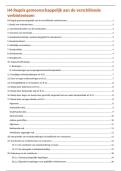
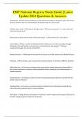
Structuren die we kunnen zien:
- I = iris
- Can’t= ciliary body band (aanhechting iris aan corpus ciliare)
- See = scleraal spoor (witte band)
- Till = trabeculair systeem (ATM en PTM = de afvoer)
- Schwalbers line = overgang tussen cornea en de afvoer. Dus het einde van descemet
Schwalbe’s lijn:
- Kleur varieert van helder (wit/ geel tot lichtbruin) vanwege de pigmentatie
- Maak optic section, verlichtingssysteem 5 tot 10 graden
- Kijk naar 2 reflecties
, 1. Reflex in de kamerhoek
2. Reflex cornea/ sclera
- Schwalbe’s lijn met pigment wordt Sampaolesi lijn
Kanaal van schlemm:
- Circulair veneus kanaal (ligt in sclera net achter/ onder het limbus
gebied)
- Bevat collector kanaaltjes voor de kamerwater afvoer
- Is soms een donkere lijn achter het trabekelsysteem
- 90% van de kamerwater afvoer verlaat het oog op deze manier
- Soms rood van kleur in schlemm’s kanaal, dan druk je te hard op de
gonio!
Trabekelsysteem (ATM en PTM):
- Bestaat uit 2 delen: anterior en posterior
- Zeef structuur: kan vol raken
- Tussen Schwalbes lijn en scleraal spoor
- Drainage (90% van de afvoer)
- Alleen posteriode deel is functioneel
- Gepigmenteerd (komt naarmate men ouder wordt)
- Naast scleraal spoor
- Grijs/ bruin doorzichtig
Scleraal spoor:
- Ring collageen en elastisch weefsel
- Het einde van trabekel systeem
- Sclerale sulcus is het herkenningspunt (driehoekje)
- Verbinding tussen sclera en longitudinale spier van het ciliary
lichaam (ciliary body)
- Witte lijn
- Moeilijk zichtbaar in lichte ogen
Ciliar lichaam:
- Productie kamerwater
- Accommodatie
- Aanhechtingspunt voor iris
- Grootste deel ligt achter de iris (niet zichtbaar)
- Zichtbaar is de ciliair lichaam band
- Grootte van de ciliair lichaam band hangt af van:
o Insertie (aanhechting) iris
o Myoop/ hypermetroop (oog bij myoop
kan groter en langer zijn
- Kleur: bruin/ grijze band (hangt af van de
pigmentatie)