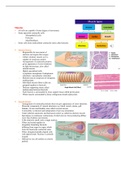
Muscles
= All cells are capable of some degree of movement.
= Some specialist contractile cells:
Myoepithelial cells
Pericytes
Myofibroblasts
= Some cells form multicellular contractile units called muscles.
a) Skeletal Muscle
o Responsible for movement of
skeleton and organs like tongue.
o Called voluntary muscle as it is
capable of conscious control.
o Arrangement of contractile proteins
gives appearance of cross-striations
on light microscopy, also called
striated muscle.
o Highly specialised cells.
Cytoplasm=sarcoplasm. Endoplasmic
reticulum= sarcoplasmic reticulum.
o Skeletal muscle composed of elongated,
multinucleate.
o Individual muscle fibres (cells) are
grouped together in fasciculi.
o Delicate supporting tissue called
endomysium between fasciculi.
o Each fascicle is surrounded by loose support tissue called perimysium.
o Whole muscle surrounded by dense collagenous sheath-epimysium.
b) Smooth Muscle
o Arrangement of contractile proteins does not give appearance of cross-striations.
o Muscular component of visceral structures e.g. blood vessels, uterus, gall
bladder, GI tract and bladder, also called visceral muscle.
o Contractility is inherent and independent of nervous stimulation.
o Under inherent autonomic and hormonal control so called involuntary muscle.
o Specialises in continuous contractions of relatively low force producing diffuse
wave-like rhythmic movements.
o Cells relatively small, only a single nucleus.
o Fibres are bound together in
irregularly branching fasciculi,
differing from organ to organ, which
form the functional contractile units.
o Fibres elongated spindle-shaped cells
with tapered ends. Nucleus is centrally
located.
o Difficult to see cell outlines as closely
packed.
, c) Cardiac Muscle
o Muscle of the myocardium.
o Many structural and functional characteristics intermediate between
skeletal and smooth muscle.
o Like skeletal muscle, contractions are strong and utilise much energy.
o Like smooth muscle, contractions are continuous and imitated by
inherent mechanisms-modulated by nerves and hormones.
o Cardiac muscle fibres are long and cylindrical, with one or 2
centrally placed nuclei.
o The ends of the cell are split longitudinally and about on to similar
branches on adjacent cells.
o Between fibres, equivalent of endomysium is seen, supports rich
capillary network, necessary to meet the high metabolic demands of
strong, continuous activity.
o Arrangement of contractile proteins similar to skeletal muscle,
striations less obvious as myofibrils and cells are more irregularly
shaped.
o Elongated nuclei mainly centrally located.
o Contraction of myocardium as a whole in each cardiac cycle is coordinated by highly modified cardiac
cells= Purkinje system.
Skeletal System: Bone
- Bone ▪ The primary tissue of bone= osseous tissue.
- Cartilage ▪ Hard and light weight composite material. Mostly calcium
- Joints phosphate as calcium hydroxyapatite.
- Tendons ▪ High compressive strength but poor tensile strength. Degree of
- Ligaments elasticity, contributed chiefly by collagen.
▪ All bones consists of living and dead cells embedded in the
mineralized organic matrix that makes up the osseous tissue.
Bone Structure:
Central Haversian Canals: run longitudinally within the substance of the bone and contain the
blood vessels, which nourish the osteocytes.
Central canals are surrounded by 4 to 20 successive layers of bone known as the concentric
lamellae. Each canal and its associated concentric lamellae constitute a unit of bone structure
called Haversian system or osteone.
Bone Cell Types
Osteoblasts→ synthesize osteoid and mineralise it. Link up on surface of bone.
Osteocytes→ inactive osteoblasts. Maintain nutrition of bone.
Osteoclasts→ phagocytic cells. Involved in bone turnover.
Two types of bone can be identified microscopically according tot the pattern of collagen
forming the osteoid (collagenous support tissue of type I collagen embedded in
glycosaminoglycan gel):
1) Woven bone: characterised by haphaza and organisation of collagen
fibres and is mechanically weak. Disorganized collagen fibres.
Immature, produced rapidly. Fracture.
2) Lamellar bone: which has a regular parallel alignment of
collagen into sheets (lamellae) and is mechanically strong.
Parallel bands of collagen fibres. Mature, produced gradually
via osteoclastic remodelling.
, Joints
a) Synovial→ extensive movement.
Articular surfaces coated
with synovial fluid.
b) Non-synovial→ no
free articulating surface.
Sutures of skull bones
(syndesmoses). 1st rib
with sternum
(synchondroses).
Intervertebral discs (fibrocartilage joints-sympheses).
Synovium
- Synovial cells of mesenchymal origin.
- No basement membrane
- No cell-cell junctions.
Tendon
Attaches skeletal muscle to bones.
Capable of withstanding tension.
Transmit forces.
Some tendons can store energy and acts as spring to make
locomotion more efficient.
Ligament
Connect bones to bones at joints.
Also formed of dense regular connective tissue.
Greater proportion of elastic fibres than tendons.
Limited regenerative capacity.
Female and Male Reproductive System
Female Reproductive System Functions:
• Production of female gametes, the ova, by the process of oogenesis.
• Reception of male gametes, the spermatozoa.
• Provision of a suitable environment for the fertilization of ova by spermatozoa.
• Expulsion of the developed foetus to the external environment.
• Nutrition of the new-born.
• Structure:
Ovaries→ site of oogenesis. Paired organs lying either side of the uterus. Ova are
released in a cyclical manner (ovulation). Produce oestrogen and progesterone.
Genitelia Tract→ provides environment for: reception of male gametes, fertilisation
of ova, development and expulsion of the foetus.
Breasts→ during pregnancy, the secretory components expand greatly in size and
number in preparation for milk production (lactation).