3.6 BRAIN ANATOMY
LEARNING GOALS
During this practical you will learn about the anatomy of the human brain. At the end of this
practical you will have profound knowledge of the anatomy of the human brain. You will be
able to identify and describe:
- the four cerebral lobes and the main gyri and sulci of the lobes
- the structures on the midsagittal plane
- the white matter tracts in the brain
- the structures that belong to the ventricular system
- the structures that belong to the limbic system
- the structures that belong to the basal ganglia
- the structures of the diencephalon
- the structures of the brain stem and the cerebellum
- the layers that protect the brain from the outside
WORKBOOK 1: OUTSIDE THE BRAIN
Use program BrainVoyager Brain Tutor
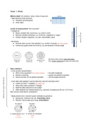
ORIENTATION
Dorsal = toward the side of the back
Ventral = toward the side of the belly
Anterior = toward the front
,Posterior = toward the back
Medial = toward the midline
Lateral = away from the midline
Superior = in the direction of the head
Inferior = in the direction of the feet
Proximal = close by
Distal = far away
Ipsilateral = at the same side
Contralateral = at the other side
Rostral = toward the direction of the beak/snout
Caudal = toward the direction of the tail
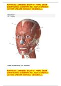
LOBES
!Note that there are individual differences in the pattern of sulci and gyri between two brains,
although the rough pattern is similar in all individuals
● Frontal lobe is at the front and is the largest lobe, involved in executive functions and
motor control
● Parietal lobe is at the upper part and is involved in perception and sensory
processing
● Temporal lobe is at the side and is involved with memory, understanding and
language
● Occipital lobe is at the back and is involved in vision
● At the midsagittal view you can see all four lobes
● In the top view, only a small portion of the occipital lobe can be seen
● The border between frontal and parietal lobe is the central sulcus
● The border between occipital and parietal lobe is hardly determined from a lateral
view, but more likely seen in a midsagittal section
● Poles refer to endings or most extreme points of the lobes
, ● Determine front and back of the brain from top view: 3 horizontal lines (superior,
medial and inferior gyri) pertain to the frontal lobe, occipital lobe is barely seen
LEARNING GOALS
During this practical you will learn about the anatomy of the human brain. At the end of this
practical you will have profound knowledge of the anatomy of the human brain. You will be
able to identify and describe:
- the four cerebral lobes and the main gyri and sulci of the lobes
- the structures on the midsagittal plane
- the white matter tracts in the brain
- the structures that belong to the ventricular system
- the structures that belong to the limbic system
- the structures that belong to the basal ganglia
- the structures of the diencephalon
- the structures of the brain stem and the cerebellum
- the layers that protect the brain from the outside
WORKBOOK 1: OUTSIDE THE BRAIN
Use program BrainVoyager Brain Tutor
ORIENTATION
Dorsal = toward the side of the back
Ventral = toward the side of the belly
Anterior = toward the front
,Posterior = toward the back
Medial = toward the midline
Lateral = away from the midline
Superior = in the direction of the head
Inferior = in the direction of the feet
Proximal = close by
Distal = far away
Ipsilateral = at the same side
Contralateral = at the other side
Rostral = toward the direction of the beak/snout
Caudal = toward the direction of the tail
LOBES
!Note that there are individual differences in the pattern of sulci and gyri between two brains,
although the rough pattern is similar in all individuals
● Frontal lobe is at the front and is the largest lobe, involved in executive functions and
motor control
● Parietal lobe is at the upper part and is involved in perception and sensory
processing
● Temporal lobe is at the side and is involved with memory, understanding and
language
● Occipital lobe is at the back and is involved in vision
● At the midsagittal view you can see all four lobes
● In the top view, only a small portion of the occipital lobe can be seen
● The border between frontal and parietal lobe is the central sulcus
● The border between occipital and parietal lobe is hardly determined from a lateral
view, but more likely seen in a midsagittal section
● Poles refer to endings or most extreme points of the lobes
, ● Determine front and back of the brain from top view: 3 horizontal lines (superior,
medial and inferior gyri) pertain to the frontal lobe, occipital lobe is barely seen