KEY CONCEPTS Muscle tissue Organisation
▪ Muscle types
▪ Describe the differences in structure between the
three muscle types
▪ Structure of striated muscle tissue
▪ Describe the gross and microscopic structure of
striated muscle
▪ Striated muscle contraction
▪ List in sequence the events that take place during
muscle contraction
▪ Explain the sliding filament theory
▪ Control of movement
▪ Describe how muscles work, including the
antagonistic action of skeletal
▪ Muscles
- Contracts involuntarily, eg. movement of - Contraction is involuntary
food through the digestive tract
Antagonistic action of skeletal muscles
• Agonist muscle contracts
• Antagonist muscle relaxes
• Groups of muscles work together
• Separate, closely timed stimuli produce smooth,
sustained contraction
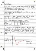
- Under voluntary control The physical interaction of protein filaments is
- Essential for locomotion required for muscle function
→ Muscle cell contraction relies on the interaction
between protein structures called thin and thick
filaments.
→ The major component of thin filaments is the
globular protein actin.
→ In thin filaments, two strands of polymerized actin
are coiled around one another; similar actin
structures called microfilaments function in cell
motility.
→ The thick filaments are staggered arrays of myosin
molecules.
→ Muscle contraction is the result of filament
movement powered by chemical energy; muscle
TJW NOTES extension occurs only passively
, Vertebrate Skeletal Muscle
Structure and Function of Vertebrate Skeletal
Muscle:
→ Vertebrate skeletal muscle, which moves bones
and body, has a hierarchy of smaller and smaller
units.
→ Within a typical skeletal muscle is a bundle of long
fibers running along the length of the muscle.
Each individual fiber is a single cell.
→ Within are multiple nuclei, each derived from one
of the embryonic cells that fused to form the fiber.
→ Surrounding these nuclei are longitudinal
myofibrils, which consist of bundles of thin and
thick filaments.
→ The myofibrils in muscle fibers are made up of
repeating sections called sarcomeres ~ which are
the basic contractile units of skeletal muscle.
→ The borders of the sarcomere line up in adjacent
myofibrils, forming a pattern of light and dark
bands (striations) visible with a light microscope.
→ Thin filaments attach at the Z lines at the
sarcomere ends
→ Thick filaments are anchored in the middle of the
sarcomere (M line).
→ In a resting (relaxed) myofibril, thick and thin
filaments partially overlap.
→ Near the edge of the sarcomere there are only
thin filaments, whereas the zone in the center
contains only thick filaments.
→ The sarcomere is the functional unit of muscle
contraction.
myofilament sacromere
TJW NOTES
▪ Muscle types
▪ Describe the differences in structure between the
three muscle types
▪ Structure of striated muscle tissue
▪ Describe the gross and microscopic structure of
striated muscle
▪ Striated muscle contraction
▪ List in sequence the events that take place during
muscle contraction
▪ Explain the sliding filament theory
▪ Control of movement
▪ Describe how muscles work, including the
antagonistic action of skeletal
▪ Muscles
- Contracts involuntarily, eg. movement of - Contraction is involuntary
food through the digestive tract
Antagonistic action of skeletal muscles
• Agonist muscle contracts
• Antagonist muscle relaxes
• Groups of muscles work together
• Separate, closely timed stimuli produce smooth,
sustained contraction
- Under voluntary control The physical interaction of protein filaments is
- Essential for locomotion required for muscle function
→ Muscle cell contraction relies on the interaction
between protein structures called thin and thick
filaments.
→ The major component of thin filaments is the
globular protein actin.
→ In thin filaments, two strands of polymerized actin
are coiled around one another; similar actin
structures called microfilaments function in cell
motility.
→ The thick filaments are staggered arrays of myosin
molecules.
→ Muscle contraction is the result of filament
movement powered by chemical energy; muscle
TJW NOTES extension occurs only passively
, Vertebrate Skeletal Muscle
Structure and Function of Vertebrate Skeletal
Muscle:
→ Vertebrate skeletal muscle, which moves bones
and body, has a hierarchy of smaller and smaller
units.
→ Within a typical skeletal muscle is a bundle of long
fibers running along the length of the muscle.
Each individual fiber is a single cell.
→ Within are multiple nuclei, each derived from one
of the embryonic cells that fused to form the fiber.
→ Surrounding these nuclei are longitudinal
myofibrils, which consist of bundles of thin and
thick filaments.
→ The myofibrils in muscle fibers are made up of
repeating sections called sarcomeres ~ which are
the basic contractile units of skeletal muscle.
→ The borders of the sarcomere line up in adjacent
myofibrils, forming a pattern of light and dark
bands (striations) visible with a light microscope.
→ Thin filaments attach at the Z lines at the
sarcomere ends
→ Thick filaments are anchored in the middle of the
sarcomere (M line).
→ In a resting (relaxed) myofibril, thick and thin
filaments partially overlap.
→ Near the edge of the sarcomere there are only
thin filaments, whereas the zone in the center
contains only thick filaments.
→ The sarcomere is the functional unit of muscle
contraction.
myofilament sacromere
TJW NOTES