Radio-anatomie- MSK
1. Syndesmologie
- Beenderen ® stevigheid
- Beenverbindingen® beweeglijkheid + stabiliteit van twee naast
elkaar gelegen beenderen
- Epifysaire schijf ® lengtegroei
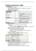
1.1 Classificatie gewrichten
Onbeweeglijke
beenverbinding
Satura Verbinding bestaat uit een zeer dunne
lijn (schedelnaad)
Synchondrosis Kraakbeen dient als tussenstof (tussen
rib-sternum)
Synostosis Beenderige vergroeiing van primitief
gescheiden botstukken (sacrum)
Synarthrosis
Amphiarthrosis Weinig beweeglijke beenverbinding
Syndesmo Tussenliggend weefsel bestaat uit
sis vlezig bindweefsel/ ligamenten (tibia-
fibula)
Symphisis Kraakbeen vormt tussenliggend
weefsel (disci intervertebrales)
Diarthrosis Beweeglijke beenverbinding
1.2. Opbouw diarthrose
- Facies articulares® bekleed met hyalien kraakbeen
- Tweeledig:
o Caput articularis (convex)
=Congruent gewichtsopp.
(Indien niet goed op elkaar = discongruentie)
o Cavitas articularis (concaaf)
- Gewricht: omgeven door capsula(compensatie)
articularis
- Binnenste laag kapsel: membrana synovialis
o Vaatrijk + uitscheiding synoviaal vocht
o Synoviaal vocht: voeding kraakbeen + smeermiddel van
bursae synovialis
- Buitenste laag kapsel: membrana fibrosa
- Ligamenten: versterken gewricht + passieve stabiliteit
o Inta- of extra capsulair zijn
1.2.1. Types gewrichtsvormen bij de diartrosis
Ginglymus Scharniergewr Éénassig
icht
Art. trochlearis Schroefgewric Éénassig
ht
Art. trochoidea Rol/ Éénassig
draaigewricht
Art. ellipsoidea Ei-gewricht Twee-assig
Art. sellaris Zadelgewricht Twee-assig
Art. spheroidea Kogelgewricht drieassig
- Glijdend gewricht: beweging naar vele richtingen in 1 enkel vlak
- Scharniergewricht: hoekbeweging in één enkel gewricht
- Draaigewricht: rotatie in één vlak
- Condylair gewricht: hoekbeweging in 2 vlakken
- Zadelgewricht: hoekbeweging in 2 vlakken en circumductie
, - Kogelgewricht: hoekbeweging, rotatie en circumductie
1.3. Topografie
- De anatomische houding:
o Rechtop staan
o Hoofd: rechtopstaande positie
o Armen gestrekt+ naast lichaam
o Handpalmen naar voor+ duimen naar buiten
o Voeten licht gespreid
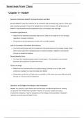
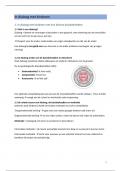
- Doorsnedes en lichaamsvlakken:
o Transversaal/
axiaal vlak®
verdeeld lichaam
in craniaal en
caudaal
^ =
lonitudinale
as
o Frontaal/
coronaal vlak ®verdeeld lichaam in anterior en posterior
^ = sagittale as
o Sagittale vlak® verdeeld lichaam in sinistra (links) en
dextra (rechts)
^= transversale as
o Mediaalvlak/ medio-sagittale vlak® strikt sagittale
doorsnede dat 2 gelijke lichaamshelften geeft
- Plaatsaanduidingen:
- Richtingsaanduidingen:
o Adductie: van de mediaanlijn af in het frontale vlak
o Abductie: naar de mediaanlijn toe in het frontale vlak
o (ante) flexie: naar ventraal in sagittale vlak
o (retroflexie) extensie: naar dorsaal in sagittale vlak
o Endorotatie: binnenwaarts draaien van botstuk/ mediale
rotatie
o Exorotatie: buitenwaarts draaien van botstuk/ laterale
rotatie
o Pronatie: handpalm naar dorsaal draaien
, o Supinatie: handpalm naar ventraal draaien
o Eversie: voetzool naar buiten draaien
o Inversie: voetzool naar binnen draaien
2. Beeldherkenning
- Projectie radiologie (2D)
o 3D volume dmv superpositie vastgelegd als 2D projectie
o Plaatsaanduiding: loodletters
- CT & MR8 (3D)
o CT matrix: 512x512
o Ruwe data + weergave adhv FOV in XY-vlak
o Reconstructive bij MSK volgens inclinatiehoek uitgevoerd
die bepaald wordt door verloop gewrichtsvlak
Richting benoemen met para- (para-sagittaal, para-
coronaal,…)
3. Het bovenste lidmaat
- 29 botstructuren
- 3 groepen:
o Ossa digitorum
manus
o Ossa metacarpi
o Ossa carpi
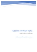
3.1. Ossa digitorum manus
- Synoniem: phalanges
- Genummerd vanaf duim nr
pink (I-V)
- 3 delen:phalanx proximalis,
phalanx media en phalanx
distalis
- Duim/ pollex: slechts
phalanx proximalis en
distalis
- Phalanx: 3 delen® caput,
basis en corpus
- Phalangis distalis:
o Palmaire zijde: ruw opp= tuberositas phalangis distalis
Sluit corpus distalis af
- Phalangis media:
o Glijdingsranden vormen gewricht met caput phalangis
proximalis
- Phalangis proximalis:
o Plantair: ruw/ concaaf® aanhechting spieren
o Dorsaal: glad en convex
o Art. interphalangae: verbinding tussen 2 phalangen
DIP: distaal interphalangaele gewrichten
PIP: proximaal interphalangaele gewrichten
o Duim: enkel interphalangeaal gewricht
Gewricht tussen metacarpi en phalanx: art.
metacarpophalangeale I
, 3.2. Ossa metacarpi
- 5 kokervormige beentjes + driehoekige doorsnede
- Palmair: sulcus carpi
- Caput, corpus en basis ossis metacarpi
- Caput ossis metacarpi en basis proximale phalanx= art.
metacarpophalangeale I-V (MCP)
o Kogelgewrichten: flexie- extensie van vinger
o Aan laterale en mediale zijde van art.
metacarpophalangeale I-V® ligg. Collateralia
ligg. Collateralia: AD- en Abductie beweging
beperken
- Cartilago articularis: vergroot contact opp. + bepaald ROM van
gewricht
- Duim: zadelvormig gewricht
o Flexie-extensie
o ADductie en Abductie
o Oppositie en repositie
- Basis ossis metacarpi en ossa carpi® art. Carpometacarpales
- Ossa metacarpale II en III: processus styloideus ossis metacarpi
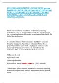
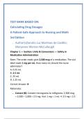
3.3. Ossa carpi
- 2 rijen van 4 beentjes
- Ossa carpi proximalia:
o Os scaphoïdeum, os lunatum, os triquetrum en os
pisiforme
- Ossa carpi distalia:
o Os trapezium, os trapezoideum, os capitatum en os
hamatum
o Os hamatum bevat hamulus ossis hamate (palmaire zijde)
- Some Lovers Try Positions That They Can’t Handle®
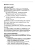
- Canalis carpi (carpal tunnel): bevat N. medianus
(carpaaltunnelsyndroom)
o Ulnaire rand gevormd door os pisiforme en hamulus ossis
hamati
o Radiale rand gevormd door tuberculum ossis scaphoidei en
tuberculum ossis trapezium
o Goot wordt overspannen met retinaculum fexorum
- Onderverdeling canalis carpi in kolommen:
o Laterale kolom: kolom van de duim
o Middelste kolom: os lunatum, os capitatum en metacarpaal
III® vormen as van de hand
o Mediale kolom: 2 laatste vingers, os triquetrum en os
hamatum, metacarpaal IV-V
1. Syndesmologie
- Beenderen ® stevigheid
- Beenverbindingen® beweeglijkheid + stabiliteit van twee naast
elkaar gelegen beenderen
- Epifysaire schijf ® lengtegroei
1.1 Classificatie gewrichten
Onbeweeglijke
beenverbinding
Satura Verbinding bestaat uit een zeer dunne
lijn (schedelnaad)
Synchondrosis Kraakbeen dient als tussenstof (tussen
rib-sternum)
Synostosis Beenderige vergroeiing van primitief
gescheiden botstukken (sacrum)
Synarthrosis
Amphiarthrosis Weinig beweeglijke beenverbinding
Syndesmo Tussenliggend weefsel bestaat uit
sis vlezig bindweefsel/ ligamenten (tibia-
fibula)
Symphisis Kraakbeen vormt tussenliggend
weefsel (disci intervertebrales)
Diarthrosis Beweeglijke beenverbinding
1.2. Opbouw diarthrose
- Facies articulares® bekleed met hyalien kraakbeen
- Tweeledig:
o Caput articularis (convex)
=Congruent gewichtsopp.
(Indien niet goed op elkaar = discongruentie)
o Cavitas articularis (concaaf)
- Gewricht: omgeven door capsula(compensatie)
articularis
- Binnenste laag kapsel: membrana synovialis
o Vaatrijk + uitscheiding synoviaal vocht
o Synoviaal vocht: voeding kraakbeen + smeermiddel van
bursae synovialis
- Buitenste laag kapsel: membrana fibrosa
- Ligamenten: versterken gewricht + passieve stabiliteit
o Inta- of extra capsulair zijn
1.2.1. Types gewrichtsvormen bij de diartrosis
Ginglymus Scharniergewr Éénassig
icht
Art. trochlearis Schroefgewric Éénassig
ht
Art. trochoidea Rol/ Éénassig
draaigewricht
Art. ellipsoidea Ei-gewricht Twee-assig
Art. sellaris Zadelgewricht Twee-assig
Art. spheroidea Kogelgewricht drieassig
- Glijdend gewricht: beweging naar vele richtingen in 1 enkel vlak
- Scharniergewricht: hoekbeweging in één enkel gewricht
- Draaigewricht: rotatie in één vlak
- Condylair gewricht: hoekbeweging in 2 vlakken
- Zadelgewricht: hoekbeweging in 2 vlakken en circumductie
, - Kogelgewricht: hoekbeweging, rotatie en circumductie
1.3. Topografie
- De anatomische houding:
o Rechtop staan
o Hoofd: rechtopstaande positie
o Armen gestrekt+ naast lichaam
o Handpalmen naar voor+ duimen naar buiten
o Voeten licht gespreid
- Doorsnedes en lichaamsvlakken:
o Transversaal/
axiaal vlak®
verdeeld lichaam
in craniaal en
caudaal
^ =
lonitudinale
as
o Frontaal/
coronaal vlak ®verdeeld lichaam in anterior en posterior
^ = sagittale as
o Sagittale vlak® verdeeld lichaam in sinistra (links) en
dextra (rechts)
^= transversale as
o Mediaalvlak/ medio-sagittale vlak® strikt sagittale
doorsnede dat 2 gelijke lichaamshelften geeft
- Plaatsaanduidingen:
- Richtingsaanduidingen:
o Adductie: van de mediaanlijn af in het frontale vlak
o Abductie: naar de mediaanlijn toe in het frontale vlak
o (ante) flexie: naar ventraal in sagittale vlak
o (retroflexie) extensie: naar dorsaal in sagittale vlak
o Endorotatie: binnenwaarts draaien van botstuk/ mediale
rotatie
o Exorotatie: buitenwaarts draaien van botstuk/ laterale
rotatie
o Pronatie: handpalm naar dorsaal draaien
, o Supinatie: handpalm naar ventraal draaien
o Eversie: voetzool naar buiten draaien
o Inversie: voetzool naar binnen draaien
2. Beeldherkenning
- Projectie radiologie (2D)
o 3D volume dmv superpositie vastgelegd als 2D projectie
o Plaatsaanduiding: loodletters
- CT & MR8 (3D)
o CT matrix: 512x512
o Ruwe data + weergave adhv FOV in XY-vlak
o Reconstructive bij MSK volgens inclinatiehoek uitgevoerd
die bepaald wordt door verloop gewrichtsvlak
Richting benoemen met para- (para-sagittaal, para-
coronaal,…)
3. Het bovenste lidmaat
- 29 botstructuren
- 3 groepen:
o Ossa digitorum
manus
o Ossa metacarpi
o Ossa carpi
3.1. Ossa digitorum manus
- Synoniem: phalanges
- Genummerd vanaf duim nr
pink (I-V)
- 3 delen:phalanx proximalis,
phalanx media en phalanx
distalis
- Duim/ pollex: slechts
phalanx proximalis en
distalis
- Phalanx: 3 delen® caput,
basis en corpus
- Phalangis distalis:
o Palmaire zijde: ruw opp= tuberositas phalangis distalis
Sluit corpus distalis af
- Phalangis media:
o Glijdingsranden vormen gewricht met caput phalangis
proximalis
- Phalangis proximalis:
o Plantair: ruw/ concaaf® aanhechting spieren
o Dorsaal: glad en convex
o Art. interphalangae: verbinding tussen 2 phalangen
DIP: distaal interphalangaele gewrichten
PIP: proximaal interphalangaele gewrichten
o Duim: enkel interphalangeaal gewricht
Gewricht tussen metacarpi en phalanx: art.
metacarpophalangeale I
, 3.2. Ossa metacarpi
- 5 kokervormige beentjes + driehoekige doorsnede
- Palmair: sulcus carpi
- Caput, corpus en basis ossis metacarpi
- Caput ossis metacarpi en basis proximale phalanx= art.
metacarpophalangeale I-V (MCP)
o Kogelgewrichten: flexie- extensie van vinger
o Aan laterale en mediale zijde van art.
metacarpophalangeale I-V® ligg. Collateralia
ligg. Collateralia: AD- en Abductie beweging
beperken
- Cartilago articularis: vergroot contact opp. + bepaald ROM van
gewricht
- Duim: zadelvormig gewricht
o Flexie-extensie
o ADductie en Abductie
o Oppositie en repositie
- Basis ossis metacarpi en ossa carpi® art. Carpometacarpales
- Ossa metacarpale II en III: processus styloideus ossis metacarpi
3.3. Ossa carpi
- 2 rijen van 4 beentjes
- Ossa carpi proximalia:
o Os scaphoïdeum, os lunatum, os triquetrum en os
pisiforme
- Ossa carpi distalia:
o Os trapezium, os trapezoideum, os capitatum en os
hamatum
o Os hamatum bevat hamulus ossis hamate (palmaire zijde)
- Some Lovers Try Positions That They Can’t Handle®
- Canalis carpi (carpal tunnel): bevat N. medianus
(carpaaltunnelsyndroom)
o Ulnaire rand gevormd door os pisiforme en hamulus ossis
hamati
o Radiale rand gevormd door tuberculum ossis scaphoidei en
tuberculum ossis trapezium
o Goot wordt overspannen met retinaculum fexorum
- Onderverdeling canalis carpi in kolommen:
o Laterale kolom: kolom van de duim
o Middelste kolom: os lunatum, os capitatum en metacarpaal
III® vormen as van de hand
o Mediale kolom: 2 laatste vingers, os triquetrum en os
hamatum, metacarpaal IV-V