NUR3065 Exam #3 – Study Guide
I. Abdomen and Gastrointestinal System
Assessment Sequence- Inspection → Auscultation → Percussion → Palpation (avoid altering bowel
sounds)
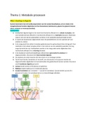
Organs Found in Each Quadrant (figure 16-3):
RUQ- LUQ-
Liver, gallbladder, right kidney, duodenum, Stomach, spleen, pancreas tail, left kidney.
ascending/transverse colon. Pain: Pancreatitis—radiates to back, worse after fatty
Pain: Gallstones (Murphy’s sign positive—pain with meals.
inspiration during RUQ palpation).
RLQ- LLQ-
Appendix, cecum, right ovary/tube. Sigmoid & descending colon left ovary/tube
Pain: Appendicitis (starts periumbilical → localizes to Pain: Diverticulitis—crampy LLQ pain, fever,
RLQ). leukocytosis.
Abdominal Pain Types (pg. 504-506)
Type Description Examples
Visceral Dull, crampy, poorly localized. From organ distention or Early appendicitis, cholecystitis, bowel obstruction.Ma
stretching.hollow abdominal organs such as the intestine be difficult to localize. It is typically palpable near the
or biliary tree contract unusually forcefully or are midline at levels that vary according to the structure
distended or stretched. involved, as illustrated in Figure 16-5. Visceral pain
varies in quality and may be gnawing, burning, crampin
or aching. When it becomes severe, it may be associate
with sweating, pallor, nausea, vomiting, and restlessnes
Parietal Sharp, localized, aggravated by movement.It is a steady, Late appendicitis, peritonitis. riginates from inflammati
aching pain that is usually more severe than visceral pain in the parietal peritoneum
and more precisely localized over the involved structure.
It is typically aggravated by movement or coughing
Referred Felt in area distant from origin due to shared Gallbladder → right shoulder (Kehr’s sign); Pancreas →
innervation.is felt in more distant sites, which are back; MI → epigastrium.
innervated at approximately the same spinal levels as the
inflamed structures. Referred pain often develops as the
initial pain becomes more intense and thus seems to
radiate or travel from the initial site. It may be palpated
superficially or deeply but is usually well localized. Pain
may also be referred to the abdomen from the chest,
spine, or pelvis, and complicate the assessment of
abdominal pain.
Common GI Disorders
Disorder Key Findings Diagnostic Signs
,Appendicitis The pain of appendicitis classically begins near Psoas sign- Place your hand just above the patient’s
( about 6 questions) the umbilicus, then migrates to the RLQ. Older right knee and ask the patient to raise that thigh again
adults are less likely to report this pattern. your hand. Alternatively, ask the patient to turn onto t
left side. Then extend the patient’s right leg at the hip
McBurney’s Point-lies 2 in from the anterior Flexion of the leg at the hip makes the psoas muscle
superior spinous process of ilium on a line contract; extension stretches it. Increased abdominal
drawn from that process to the umbilicus pain on either maneuver is a positive psoas sign,
suggesting irritation of the psoas muscle by an inflam
appendix.
Rosving’s sign- Press deeply and evenly in the left low
quadrant. Then quickly withdraw your fingers. Pain i
the RLQ during left-sided pressure is a positive Rovs
sign. Pain in the RLQ when pressure is released from
the LLQ is referred rebound tenderness.
Obturator sign-(though this sign has very low
sensitivity). Flex the patient’s right thigh at the hip wi
the knee bent and rotate the leg internally at the hip.
This maneuver stretches the internal obturator muscle
Right hypogastric pain is a positive obturator sign,
suggesting irritation of the obturator muscle by an
inflamed appendix.
Diverticulitis LLQ pain, especially with a palpable mass,
signals
iffuse abdominal pain with abdominal
distention, hyperactive high-pitched bowel
sounds, and tenderness on palpation may
indicate small or large bowel obstruction. Pain
with absent bowel sounds, rigidity, percussion
tenderness, and guarding points to peritonitis.
Triggers: often a change in bowel habits, weight
loss
Often no other symptoms Often no other
symptoms unless inflammation causes
Pancreatitis Epigastyric pain Doubling over with cramping,
colicky pain may signal a renal stone. Sudden
knife-like epigastric pain
Cholecystitis RUQ pain inflammation of the gallbladder, Murphey’s sign-A sharp increase in tenderness with
assess Murphy sign inspiratory effort is a positive Murphy sign. When
, ook your left thumb or the fingers of your right positive, Murphy sign triples the likelihood of acute
hand under the costal margin at the point where cholecystitis
the lateral border of the rectus muscle intersects
with the costal margin. Alternatively, palpate
the RUQ with the fingers of your right hand
near the costal margin. If the liver is enlarged,
hook your thumb or fingers under the liver edge
at a comparable point below. Ask the patient to
take a deep breath, which forces the liver and
gall bladder down toward the examining
fingers. Watch the patient’s breathing and note
the degree of tenderness.
Irritable Bowel termittent pain for 12 weeks of the preceding 12 Some patients may complain of passing excessive gas
Syndrome months with relief from defecation, change in or flatus, normally about 600 mL per day
frequency of bowel movements, or change in Functional change in frequency or form of bowel
form of stool (loose, watery, pellet-like) without movement without known pathology; possibly from
structural or biochemical abnormalities are change in intestinal bacteria
symptoms Three patterns: diarrhea-predominant,
constipation-predominant, or mixed. Symptoms prese
6 mo or longer and abdominal pain for 3 mo or longe
plus at least two of three features (improvement with
defecation; onset with change in stool frequency; ons
with change in stool form and appearance)
Ascites Shifting dullness,-Percuss the border of is the accumulation of fluid in the peritoneal cavity,
tympany and dullness with the patient supine, causing abdominal swelling.ay be due to protein
then ask the patient to roll onto one side . deficiency liver disease and low albumin.
Percuss and mark the borders again. In a person
without ascites, the borders between tympany
and dullness usually stay relatively constant.
positive fluid wave-Ask the patient or an
assistant to press the edges of both hands firmly
down the midline of the abdomen. This pressure
helps stop the transmission of a wave through
fat. While you tap one flank sharply with your
fingertips, feel on the opposite flank for an
impulse transmitted through the fluid, as shown
in Figure 16-28. Unfortunately, this sign is often
negative until ascites is obvious, and it is
sometimes positive in people without ascites.
In ascites, dullness shifts to the more dependent
side, whereas tympany shifts to the top.
I. Abdomen and Gastrointestinal System
Assessment Sequence- Inspection → Auscultation → Percussion → Palpation (avoid altering bowel
sounds)
Organs Found in Each Quadrant (figure 16-3):
RUQ- LUQ-
Liver, gallbladder, right kidney, duodenum, Stomach, spleen, pancreas tail, left kidney.
ascending/transverse colon. Pain: Pancreatitis—radiates to back, worse after fatty
Pain: Gallstones (Murphy’s sign positive—pain with meals.
inspiration during RUQ palpation).
RLQ- LLQ-
Appendix, cecum, right ovary/tube. Sigmoid & descending colon left ovary/tube
Pain: Appendicitis (starts periumbilical → localizes to Pain: Diverticulitis—crampy LLQ pain, fever,
RLQ). leukocytosis.
Abdominal Pain Types (pg. 504-506)
Type Description Examples
Visceral Dull, crampy, poorly localized. From organ distention or Early appendicitis, cholecystitis, bowel obstruction.Ma
stretching.hollow abdominal organs such as the intestine be difficult to localize. It is typically palpable near the
or biliary tree contract unusually forcefully or are midline at levels that vary according to the structure
distended or stretched. involved, as illustrated in Figure 16-5. Visceral pain
varies in quality and may be gnawing, burning, crampin
or aching. When it becomes severe, it may be associate
with sweating, pallor, nausea, vomiting, and restlessnes
Parietal Sharp, localized, aggravated by movement.It is a steady, Late appendicitis, peritonitis. riginates from inflammati
aching pain that is usually more severe than visceral pain in the parietal peritoneum
and more precisely localized over the involved structure.
It is typically aggravated by movement or coughing
Referred Felt in area distant from origin due to shared Gallbladder → right shoulder (Kehr’s sign); Pancreas →
innervation.is felt in more distant sites, which are back; MI → epigastrium.
innervated at approximately the same spinal levels as the
inflamed structures. Referred pain often develops as the
initial pain becomes more intense and thus seems to
radiate or travel from the initial site. It may be palpated
superficially or deeply but is usually well localized. Pain
may also be referred to the abdomen from the chest,
spine, or pelvis, and complicate the assessment of
abdominal pain.
Common GI Disorders
Disorder Key Findings Diagnostic Signs
,Appendicitis The pain of appendicitis classically begins near Psoas sign- Place your hand just above the patient’s
( about 6 questions) the umbilicus, then migrates to the RLQ. Older right knee and ask the patient to raise that thigh again
adults are less likely to report this pattern. your hand. Alternatively, ask the patient to turn onto t
left side. Then extend the patient’s right leg at the hip
McBurney’s Point-lies 2 in from the anterior Flexion of the leg at the hip makes the psoas muscle
superior spinous process of ilium on a line contract; extension stretches it. Increased abdominal
drawn from that process to the umbilicus pain on either maneuver is a positive psoas sign,
suggesting irritation of the psoas muscle by an inflam
appendix.
Rosving’s sign- Press deeply and evenly in the left low
quadrant. Then quickly withdraw your fingers. Pain i
the RLQ during left-sided pressure is a positive Rovs
sign. Pain in the RLQ when pressure is released from
the LLQ is referred rebound tenderness.
Obturator sign-(though this sign has very low
sensitivity). Flex the patient’s right thigh at the hip wi
the knee bent and rotate the leg internally at the hip.
This maneuver stretches the internal obturator muscle
Right hypogastric pain is a positive obturator sign,
suggesting irritation of the obturator muscle by an
inflamed appendix.
Diverticulitis LLQ pain, especially with a palpable mass,
signals
iffuse abdominal pain with abdominal
distention, hyperactive high-pitched bowel
sounds, and tenderness on palpation may
indicate small or large bowel obstruction. Pain
with absent bowel sounds, rigidity, percussion
tenderness, and guarding points to peritonitis.
Triggers: often a change in bowel habits, weight
loss
Often no other symptoms Often no other
symptoms unless inflammation causes
Pancreatitis Epigastyric pain Doubling over with cramping,
colicky pain may signal a renal stone. Sudden
knife-like epigastric pain
Cholecystitis RUQ pain inflammation of the gallbladder, Murphey’s sign-A sharp increase in tenderness with
assess Murphy sign inspiratory effort is a positive Murphy sign. When
, ook your left thumb or the fingers of your right positive, Murphy sign triples the likelihood of acute
hand under the costal margin at the point where cholecystitis
the lateral border of the rectus muscle intersects
with the costal margin. Alternatively, palpate
the RUQ with the fingers of your right hand
near the costal margin. If the liver is enlarged,
hook your thumb or fingers under the liver edge
at a comparable point below. Ask the patient to
take a deep breath, which forces the liver and
gall bladder down toward the examining
fingers. Watch the patient’s breathing and note
the degree of tenderness.
Irritable Bowel termittent pain for 12 weeks of the preceding 12 Some patients may complain of passing excessive gas
Syndrome months with relief from defecation, change in or flatus, normally about 600 mL per day
frequency of bowel movements, or change in Functional change in frequency or form of bowel
form of stool (loose, watery, pellet-like) without movement without known pathology; possibly from
structural or biochemical abnormalities are change in intestinal bacteria
symptoms Three patterns: diarrhea-predominant,
constipation-predominant, or mixed. Symptoms prese
6 mo or longer and abdominal pain for 3 mo or longe
plus at least two of three features (improvement with
defecation; onset with change in stool frequency; ons
with change in stool form and appearance)
Ascites Shifting dullness,-Percuss the border of is the accumulation of fluid in the peritoneal cavity,
tympany and dullness with the patient supine, causing abdominal swelling.ay be due to protein
then ask the patient to roll onto one side . deficiency liver disease and low albumin.
Percuss and mark the borders again. In a person
without ascites, the borders between tympany
and dullness usually stay relatively constant.
positive fluid wave-Ask the patient or an
assistant to press the edges of both hands firmly
down the midline of the abdomen. This pressure
helps stop the transmission of a wave through
fat. While you tap one flank sharply with your
fingertips, feel on the opposite flank for an
impulse transmitted through the fluid, as shown
in Figure 16-28. Unfortunately, this sign is often
negative until ascites is obvious, and it is
sometimes positive in people without ascites.
In ascites, dullness shifts to the more dependent
side, whereas tympany shifts to the top.