RENAL PATHOLOGY
Robbins and Cotran Patho Basis of Disease 10th Ed.
*use at your own risk
TRANS BY: MA.AC.CE.CC.AD.SM
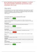
● study of kidney diseases by dividing them into those that affect ● Acute kidney injury STRUCTURES OF GLOMERULUS
the four basic morphologic components: glomeruli, tubules, ○ previously called acute renal failure Glomerular filtration barrier
interstitium, and blood vessels ○ rapid decline in GFR (within hours to days) with concurrent fluid and electrolyte imbalance, - allow discrimination among various protein molecules, depending on
● early manifestations of disease affecting each of these and retention of metabolic waste products including urea and creatinine their size ( larger, less permeable) and charge ( more cationic, the
components tend to be distinct ○ most severe forms: manifested by oliguria or anuria (reduced or no urine flow) more permeable)
● most glomerular diseases are immunologically mediated ○ can result from glomerular, interstitial, vascular, or acute tubular injury (ATI) - accounted for by the structure of the capillary wall
● tubular and interstitial disorders are frequently caused by toxic or ● Chronic kidney disease - complete exclusion of albumin, an anionic molecule
infectious agents ○ previously called chronic renal failure
● all forms of chronic kidney disease ultimately damage all four ○ presence of a diminished GFR that is persistently less than 60 mL/min/1.73 m2 for at least 3 Glomerular capillary wall is the filtering membrane and consists of the
components of the kidney, culminating end-stage kidneys months, any cause, and/or persistent albuminuria following structures:
○ end result of all chronic renal parenchymal diseases ● Endothelial cells
● End-stage renal disease ○ thin layer, fenestrated
CLINICAL MANIFESTATIONS OF RENAL DISEASES ○ GFR is less than 5% ○ 70 to 100 nm in diameter
● Azotemia: ○ terminal stage of uremia ● Glomerular basement membrane (GBM)
○ elevation of blood urea nitrogen (BUN) and creatinine ● Renal tubular defects ○ Lamina densa: thick electron-dense central layer
○ related largely to a decreased GFR ○ dominated by polyuria, , nocturia, and electrolyte disorders ○ Lamina rara interna and Lamina rara externa: thinner electron-
○ typical feature of both acute and chronic kidney injury ○ result of diseases that either directly affect tubular structures (e.g., the nephronophthisis) or lucent peripheral layers
○ Prerenal azotemia: hypoperfusion of the kidneys that impairs cause defects in specific tubular functions ○ consists of collagen (mostly type IV), laminin, polyanionic
renal function in the absence of parenchymal damage ○ can be inherited (e.g., familial nephrogenic diabetes, cystinuria, renal tubular acidosis) or proteoglycans (mostly heparan sulfate), fibronectin, entactin, and
○ Postrenal azotemia: urine flow is obstructed distal to the kidney acquired (e.g., lead nephropathy) other glycoproteins
● Uremia ● Urinary tract obstruction and renal tumors ○ Type IV Collagen: NC1 domain is important for helix formation and
○ when azotemia leads to clinical signs and symptoms ○ varied clinical manifestations for assembly of collagen monomers into the basement membrane
○ characterized not only by failure of renal excretory function but ○ based on specific location and nature suprastructure
also by metabolic and endocrine alterations from renal ○ Urinary tract infection: bacteriuria and pyuria, may be symptomatic or asymptomatic, and ● Visceral epithelial cells (podocytes)
damage may affect the kidney (pyelonephritis) or the bladder (cystitis) ○ interdigitating processes embedded in and adherent to the lamina
○ frequently manifest secondary involvement of the ● Nephrolithiasis (renal stones) rara externa
gastrointestinal system, peripheral nerves, and heart ○ spasms of severe pain (renal colic) and hematuria, with recurrent stone formation ○ Adjacent foot processes are separated by 20- to 30-nm–wide
● Nephritic syndrome filtration slits, bridged by a thin diaphragm
○ caused by inflammatory glomerular disease and dominated by ○ important for the maintenance of glomerular barrier function
the acute onset of either grossly visible hematuria or GLOMERULAR DISEASES ○ slit diaphragm presents a size-selective distal diffusion barrier to the
microscopic hematuria with dysmorphic red cells and red cell ● histologic changes in primary and secondary forms can be similar filtration of proteins
casts on urinalysis, diminished GFR, mild to moderate ● Secondary glomerular disease ○ largely responsible for synthesis of GBM components
proteinuria, and hypertension ○ Systemic immunologic diseases such as SLE, vascular disorders such as hypertension, ○ Nephrin: transmembrane protein with a large extracellular portion
○ Rapidly progressive glomerulonephritis: form of nephritic metabolic diseases such as diabetes mellitus, and some hereditary conditions such as Fabry made up of immunoglobulin (Ig)-like domains, forms molecular
syndrome, rapid decline in GFR (within hours to days) disease connections with podocin, CD2-associated protein, and ultimately
● Nephrotic syndrome ● Primary glomerulonephritis the actin cytoskeleton of the visceral epithelial cells
○ heavy proteinuria (more than 3.5 g/ day), hypoalbuminemia, ○ kidney is the only or predominant organ involved
severe edema, hyperlipidemia, and lipiduria ● Primary glomerulopathy
● Asymptomatic hematuria or proteinuria ○ do not have cellular inflammatory component
○ usually a manifestation of subtle or mild glomerular
abnormalities
PATHOLOGIC RESPONSES OF THE GLOMERULUS TO INJURY PATHOGENESIS OF GLOMERULAR INJURY MEDIATORS OF GLOMERULAR INJURY
● Hypercellularity ● Immune mechanisms underlie most forms of primary glomerulopathy and many secondary ● Cells
- increase in the number of cells in the glomerular tufts glomerular disorders ○ Neutrophils and Monocytes
- results from one or more of the following: ● Two forms of antibody-associated injury by: ■ infiltrate glomeruli in certain types of glomerulonephritis
○ Proliferation of mesangial or endothelial cells ○ Antibodies reacting in situ within the glomerulus, either binding to insoluble fixed (intrinsic) ■ result of activation of complement, resulting in generation of
○ Infiltration of leukocytes: neutrophils, monocytes, and, in some glomerular antigens or extrinsic molecules planted within the glomerulus chemotactic agents (mainly C5a), but also by Fc receptor–
1
, diseases, lymphocytes. combination of infiltration of leukocytes ○ deposition of circulating antigen-antibody complexes in the glomerulus mediated activation
and swelling and proliferation of mesangial and/or endothelial ● Major cause of glomerulonephritis resulting from formation of antigen-antibody complexes is ■ Neutrophils release proteases, cause GBM degradation;
cells (endocapillary proliferation) the consequence of in situ immune complex formation, and not deposition of circulating oxygen-derived free radicals, cause cell damage; and
○ Formation of crescents: accumulations of cells composed of complexes arachidonic acid metabolites, contribute to the reductions in
proliferating glomerular epithelial cells and infiltrating GFR
leukocytes, occurs following an immune/inflammatory injury ○ Macrophages and T lymphocytes
involving the capillary walls. Activation of coagulation factors, DISEASES CAUSED BY IN SITU FORMATION OF IMMUNE COMPLEXES ■ infiltrate glomeruli in antibody- and cell-mediated reactions
thrombin, is suspected of being a trigger for crescent formation ● Immune complexes are formed locally by antibodies that react with intrinsic tissue antigens or ○ Platelets
● Basement Membrane Thickening with extrinsic antigens “planted” in the glomerulus ■ aggregate in glomeruli during immune-mediated injury
○ Light microscopy: thickening of capillary walls, best seen in ● Membranous nephropathy is the classic example ■ release of eicosanoids, growth factors, and other mediators may
sections stained with periodic acid–Schiff (PAS) ○ Pattern of immune deposition by Immunofluorescence microscopy is granular contribute to vascular injury and proliferation of glomerular cells
○ Electron microscopy, takes one of three forms: ○ Electron microscopy: presence of numerous discrete subepithelial electron-dense deposits ○ Resident glomerular cells
■ Deposition of amorphous electron-dense material, most (made up of immune reactants) ■ Mesangial cells
often immune complexes on the endothelial or epithelial ■ stimulated to produce inflammatory mediators, including
side of GBM or within the GBM reactive oxygen species (ROS), cytokines, chemokines, growth
■ Increased synthesis of the protein components of GBM, like DISEASE CAUSED BY ANTIBODIES DIRECTED AGAINST NORMAL COMPONENTS OF THE GLOMERULAR factors, eicosanoids, nitric oxide, and endothelin
in diabetic glomerulosclerosis BASEMENT MEMBRANE ■ may initiate inflammatory responses in glomeruli, even in the
■ Additional layers of GBM matrices ● Antibodies bind to intrinsic antigens along the entire length of GBM, resulting in a diffuse linear absence of leukocytic infiltration
● Hyalinosis and Sclerosis pattern of staining for the antibodies by immunofluorescence techniques ● Soluble Mediators
○ Hyalinosis ● Anti-GBM antibody–induced glomerulonephritis: less than 5% of cases of human ○ Complement activation
■ accumulation of material that is homogeneous and glomerulonephritis but severe necrotizing and crescentic glomerular damage and the clinical ■ leads to the generation of chemotactic products that induce
eosinophilic by light microscopy. syndrome of RPGN leukocyte influx (complement-neutrophil–dependent injury) and
■ Hyalin: extracellular, amorphous material composed of the formation of C5b–C9, the membrane attack complex
plasma proteins ■ C5b–C9 causes cell lysis but, in addition, it may stimulate
■ deposits may obliterate the capillary lumens of the GLOMERULONEPHRITIS RESULTING FROM DEPOSITION OF CIRCULATING IMMUNE COMPLEXES mesangial cells to produce oxidants, proteases, and other
glomerular tuft ● Glomerular injury caused by the trapping of circulating antigen-antibody complexes within mediators. Thus, even in the absence of neutrophils, C5b–C9 can
■ usually a consequence of endothelial or capillary wall injury glomeruli cause proteinuria, as has been demonstrated in experimental
■ typically the end result of various forms of glomerular ● Antibodies have no immunologic specificity for glomerulus, and the complexes localize membranous glomerulopathy.
damage because of physicochemical properties and hemodynamic factors ■ In some diseases, collectively called C3 glomerulopathies, there
○ Sclerosis ● Antigens that trigger the formation of immune complexes may be of endogenous, as in with is evidence of complement activation resulting not from
■ deposition of extracellular collagenous matrix SLE or in IgA nephropathy, or they may be exogenous, as in certain infection like antibody or immune complex deposition but from defective
■ may be confined to mesangial areas, capillary loops, or streptococcal proteins, hepatitis B, hepatitis C, Treponema pallidum, Plasmodium falciparum, regulation of the complement system.
both and several viruses, and tumor antigens
■ may result in obliteration of some or all of the capillary ● many cases antigen is unknown ○ Eicosanoids, nitric oxide, angiotensin, and endothelin - involved in
lumens the hemodynamic changes
○ Cytokines (IL-1 and TNF) - produced by infiltrating leukocytes and
● Many primary glomerulopathies are classified by their histology CELL-MEDIATED IMMUNITY IN GLOMERULONEPHRITIS resident glomerular cells, induce leukocyte adhesion and a variety
○ Diffuse: involving all of the glomeruli in the kidney ● Sensitized T cells cause glomerular injury and are involved in the progression of some of other effects
○ Global: entirety of individual glomeruli glomerulonephritides ○ Chemokines (Monocyte chemoattractant protein 1) - promote
○ Focal: only a fraction of the glomeruli ● proof of glomerulonephritis in humans resulting primarily from T-cell activation remains lacking monocyte and lymphocyte influx
○ Segmental: part of each glomerulus ○ Growth Factors (Platelet-derived growth factor) - involved in
○ Capillary loop or mesangial: predominantly capillary or mesangial cell proliferation.
mesangial regions ACTIVATION OF ALTERNATIVE COMPLEMENT PATHWAY ○ Coagulation system - also a mediator of glomerular damage
● Occurs in Dense-deposit disease, recently referred to as MPGN type II, and in C3 ● Fibrin is frequently present in the glomeruli and Bowman space
glomerulopathies in glomerulonephritis, indicative of coagulation cascade
activation, and activated coagulation factors, particularly
thrombin, may be a stimulus for crescent formation.
2
, EPITHELIAL CELL INJURY
● Podocyte injury - common to many forms of both primary and
secondary glomerular diseases, of both immune and nonimmune
etiologies
● Podocytopathy - diseases with disparate etiologies whose
principal manifestation is injury to podocytes
● GLOMERULOSCLEROSIS
● can be induced by antibodies to podocyte antigens; by
● Sclerosis involving portions of some glomeruli (also referred to as secondary FSGS
toxins, as in an experimental model of proteinuria induced
develops after many types of renal injury and can lead to proteinuria and increasing
by puromycin aminonucleoside
functional impairment
● conceivably by certain cytokines
● Glomerular sclerosis may be seen even in cases in which the primary disease was
● by certain viral infections such as human
nonglomerular.
immunodeficiency virus (HIV)
● or by incompletely characterized circulating
● TUBULAR INJURY AND INTERSTITIAL FIBROSIS
factors,postulated minimal change disease and focal
● manifested by tubular damage and interstitial inflammation, is a component of many
segmental glomerulosclerosis (FSGS)
acute and chronic glomerulonephritides.
● Loss of podocytes, which have only a very limited capacity for
● Tubulointerstitial fibrosis contributes to progression in both immune and nonimmune
replication and repair, may be a feature of multiple types of
glomerular diseases, for example, diabetic nephropathy
glomerular injury, including FSGS and diabetic nephropathy
● there is often a much better correlation of decline in renal function with the extent of
tubulointerstitial damage than with the severity of glomerular injury
MECHANISMS OF PROGRESSION IN GLOMERULAR DISEASES
● The outcome of such injury depends on several factors, including
the severity of renal damage, the nature and persistence of the
antigens, and the immune status, age, and genetic predisposition
of the host
DISEASE DESCRIPTION PATHOGENESIS MORPHOLOGY CLINICAL FEATURES TREATMENT
NEPHRITIC SYNDROME
- often characterized by inflammation in the glomeruli
- nephritic patient usually presents with hematuria, red cell casts in the urine, azotemia, oliguria, and mild to moderate hypertension
- Proteinuria and edema are common, but these are not as severe as those encountered in the nephrotic syndrome
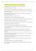
ACUTE PROLIFERATIVE - most common underlying infections - caused by immune complexes containing - diffuse proliferation of glomerular cells YOUNG CHILDREN - More than 95% of affected
(Postinfectious and are streptococcal, but the disorder streptococcal antigens and specific associated with influx (exudation) of leukocytes, - abruptly develops malaise, fever, children eventually recover
Infection-Associated) may also be associated with other antibodies typically caused by immune complexes nausea, oliguria, and hematuria renal function with conservative
GLOMERULONEPHRITIS infections - Only certain strains of group A β-hemolytic - enlarged, hypercellular glomeruli (smoky or cola-colored urine) 1 to 2
therapy aimed at maintaining
- usually appears 1 to 4 weeks after a streptococci are nephritogenic, more than - hypercellularity is caused by: weeks after recovery from a sore
streptococcal infection of the 90% of cases being traced to types 12, 4, throat. sodium and water balance
● infiltration by leukocytes, both
pharynx or skin (impetigo) and 1, which can be identified by typing of - have dysmorphic red cells or red - A small minority of children
neutrophils and monocytes
- Skin infections are commonly the M protein of the bacterial cell walls ● proliferation of endothelial and cell casts in the urine,mild proteinuria (perhaps less than 1%) do not
associated with overcrowding and - Many lines of evidence support an mesangial cells (usually less than1 g/day),periorbital improve, become severely
poor hygiene. immunologic basis for poststreptococcal ● in severe cases by crescent formation edema, and mild to moderate oliguric, and develop a rapidly
- Poststreptococcal glomerulonephritis. The latent period - There is also swelling of endothelial cells, and the hypertension progressive form of
glomerulonephritis occurs most between infection and onset of nephritis is combination of proliferation, swelling, and glomerulonephritis
frequently in children 6 to 10 years of compatible with the time required for the leukocyte infiltration obliterates the capillary ADULTS
age, but children and adults of any production of antibodies and the formation - sudden appearance of - In adults the disease is less
lumens
age can also be affected. of immune complexes. hypertension or edema benign.
- There may be interstitial edema and
- Elevated titers of antibodies against one or inflammation, and the tubules often contain red - frequently with elevation of BUN - the overall prognosis in
more streptococcal antigens are present in cell casts epidemics is good, in only
3
, a great majority of patients - immunofluorescence microscopy: there are about 60% of sporadic cases do
- The streptococcal antigenic component granular deposits of IgG and C3, and sometimes the patients recover promptly
responsible for the immune reaction had IgM in the mesangium and along the GBM - In the remainder, the
long eluded identification, but the - electron microscopic findings are discrete, glomerular lesions fail to resolve
preponderance of evidence identifies amorphous, electron-dense deposits on the
streptococcal pyogenic exotoxin B (SpeB) quickly, as manifested by
epithelial side of the membrane, often having the
as the principal antigenic determinant in appearance of “humps” presumably representing persistent proteinuria,
most but not all cases of poststreptococcal the antigen-antibody complexes at the hematuria, and hypertension
glomerulonephritis. subepithelial cell surface
- This protein can directly activate
complement, is commonly secreted by
nephritogenic strains of streptococci, and
has been localized to the “humplike”
deposits characteristic of this disease
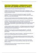
CRESCENTIC (Rapidly - a clinical syndrome associated with - a manifestation of a number of different - presence of crescents in most of the glomeruli - hematuria with red blood cell casts - Serum analyses for anti-GBM
Progressive) severe glomerular injury, but it does not diseases, some restricted to the kidney and - kidneys are enlarged and pale, often with in the urine antibodies, antinuclear
GLOMERULONEPHRITIS denote a specific etiology. others systemic petechial hemorrhages on the cortical surfaces - moderate proteinuria occasionally antibodies,andANCAsarehelpfu
- relatively rapid and progressive loss of - Although no single mechanism can - Segmental glomerular necrosis and distinctive reaching the nephrotic range
l the diagnosis of specific
renal function associated with severe explain all cases, there is little doubt that in crescents adjacent to glomerular segments - variable hypertension and edema.
oliguria and signs of nephritic most cases the glomerular injury is - Goodpasture syndrome, the course subtypes.
uninvolved by inflammatory or proliferative
syndrome immunologically mediated. changes is the feature most typical of pauci- may be dominated by recurrent - Recovery of renal function
immune RPGN hemoptysis or even life-threatening may follow early intensive
THREE GROUPS: - Fibrin strands are frequently prominent between pulmonary hemorrhage plasmapheresis (plasma
Anti-GBM antibody–mediated disease the cellular layers in the crescents exchange) combined with
- linear deposits of IgG and, in many cases, - Regardless of type, electron microscopy may steroids and cytotoxic agents in
C3 in the GBM show ruptures in the GBM, a severe injury that Goodpasture syndrome.
allows leukocytes, plasma proteins such as - Despite therapy, many
Diseases caused by immune complex coagulation factors and complement, and
deposition, with granular deposits of patients eventually require
inflammatory mediators to reach the urinary
antibodies and complement by space, where they trigger crescent formation chronic dialysis or
immunofluorescence. transplantation, particularly if
the disease is discovered at a
Pauci-immune crescentic GN, defined by late stage.
the lack of detectable anti-GBM antibodies
or immune complexes by
immunofluorescence and electron
microscopy
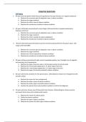
NEPHROTIC SYNDROME - caused by a derangement in - In children younger than 17 years of age in - Massive proteinuria, with the daily
glomerular capillary walls resulting in North America, for example, nephrotic loss of 3.5 g or more of protein (less in
increased permeability to plasma syndrome is almost always caused by a children)
proteins lesion primary to the kidney; among adults, - Hypoalbuminemia, with plasma
in contrast, it is often associated with a albumin levels less than 3 g/dL
systemic disease. - Generalized edema
- Hyperlipidemia and lipiduria
4
Robbins and Cotran Patho Basis of Disease 10th Ed.
*use at your own risk
TRANS BY: MA.AC.CE.CC.AD.SM
● study of kidney diseases by dividing them into those that affect ● Acute kidney injury STRUCTURES OF GLOMERULUS
the four basic morphologic components: glomeruli, tubules, ○ previously called acute renal failure Glomerular filtration barrier
interstitium, and blood vessels ○ rapid decline in GFR (within hours to days) with concurrent fluid and electrolyte imbalance, - allow discrimination among various protein molecules, depending on
● early manifestations of disease affecting each of these and retention of metabolic waste products including urea and creatinine their size ( larger, less permeable) and charge ( more cationic, the
components tend to be distinct ○ most severe forms: manifested by oliguria or anuria (reduced or no urine flow) more permeable)
● most glomerular diseases are immunologically mediated ○ can result from glomerular, interstitial, vascular, or acute tubular injury (ATI) - accounted for by the structure of the capillary wall
● tubular and interstitial disorders are frequently caused by toxic or ● Chronic kidney disease - complete exclusion of albumin, an anionic molecule
infectious agents ○ previously called chronic renal failure
● all forms of chronic kidney disease ultimately damage all four ○ presence of a diminished GFR that is persistently less than 60 mL/min/1.73 m2 for at least 3 Glomerular capillary wall is the filtering membrane and consists of the
components of the kidney, culminating end-stage kidneys months, any cause, and/or persistent albuminuria following structures:
○ end result of all chronic renal parenchymal diseases ● Endothelial cells
● End-stage renal disease ○ thin layer, fenestrated
CLINICAL MANIFESTATIONS OF RENAL DISEASES ○ GFR is less than 5% ○ 70 to 100 nm in diameter
● Azotemia: ○ terminal stage of uremia ● Glomerular basement membrane (GBM)
○ elevation of blood urea nitrogen (BUN) and creatinine ● Renal tubular defects ○ Lamina densa: thick electron-dense central layer
○ related largely to a decreased GFR ○ dominated by polyuria, , nocturia, and electrolyte disorders ○ Lamina rara interna and Lamina rara externa: thinner electron-
○ typical feature of both acute and chronic kidney injury ○ result of diseases that either directly affect tubular structures (e.g., the nephronophthisis) or lucent peripheral layers
○ Prerenal azotemia: hypoperfusion of the kidneys that impairs cause defects in specific tubular functions ○ consists of collagen (mostly type IV), laminin, polyanionic
renal function in the absence of parenchymal damage ○ can be inherited (e.g., familial nephrogenic diabetes, cystinuria, renal tubular acidosis) or proteoglycans (mostly heparan sulfate), fibronectin, entactin, and
○ Postrenal azotemia: urine flow is obstructed distal to the kidney acquired (e.g., lead nephropathy) other glycoproteins
● Uremia ● Urinary tract obstruction and renal tumors ○ Type IV Collagen: NC1 domain is important for helix formation and
○ when azotemia leads to clinical signs and symptoms ○ varied clinical manifestations for assembly of collagen monomers into the basement membrane
○ characterized not only by failure of renal excretory function but ○ based on specific location and nature suprastructure
also by metabolic and endocrine alterations from renal ○ Urinary tract infection: bacteriuria and pyuria, may be symptomatic or asymptomatic, and ● Visceral epithelial cells (podocytes)
damage may affect the kidney (pyelonephritis) or the bladder (cystitis) ○ interdigitating processes embedded in and adherent to the lamina
○ frequently manifest secondary involvement of the ● Nephrolithiasis (renal stones) rara externa
gastrointestinal system, peripheral nerves, and heart ○ spasms of severe pain (renal colic) and hematuria, with recurrent stone formation ○ Adjacent foot processes are separated by 20- to 30-nm–wide
● Nephritic syndrome filtration slits, bridged by a thin diaphragm
○ caused by inflammatory glomerular disease and dominated by ○ important for the maintenance of glomerular barrier function
the acute onset of either grossly visible hematuria or GLOMERULAR DISEASES ○ slit diaphragm presents a size-selective distal diffusion barrier to the
microscopic hematuria with dysmorphic red cells and red cell ● histologic changes in primary and secondary forms can be similar filtration of proteins
casts on urinalysis, diminished GFR, mild to moderate ● Secondary glomerular disease ○ largely responsible for synthesis of GBM components
proteinuria, and hypertension ○ Systemic immunologic diseases such as SLE, vascular disorders such as hypertension, ○ Nephrin: transmembrane protein with a large extracellular portion
○ Rapidly progressive glomerulonephritis: form of nephritic metabolic diseases such as diabetes mellitus, and some hereditary conditions such as Fabry made up of immunoglobulin (Ig)-like domains, forms molecular
syndrome, rapid decline in GFR (within hours to days) disease connections with podocin, CD2-associated protein, and ultimately
● Nephrotic syndrome ● Primary glomerulonephritis the actin cytoskeleton of the visceral epithelial cells
○ heavy proteinuria (more than 3.5 g/ day), hypoalbuminemia, ○ kidney is the only or predominant organ involved
severe edema, hyperlipidemia, and lipiduria ● Primary glomerulopathy
● Asymptomatic hematuria or proteinuria ○ do not have cellular inflammatory component
○ usually a manifestation of subtle or mild glomerular
abnormalities
PATHOLOGIC RESPONSES OF THE GLOMERULUS TO INJURY PATHOGENESIS OF GLOMERULAR INJURY MEDIATORS OF GLOMERULAR INJURY
● Hypercellularity ● Immune mechanisms underlie most forms of primary glomerulopathy and many secondary ● Cells
- increase in the number of cells in the glomerular tufts glomerular disorders ○ Neutrophils and Monocytes
- results from one or more of the following: ● Two forms of antibody-associated injury by: ■ infiltrate glomeruli in certain types of glomerulonephritis
○ Proliferation of mesangial or endothelial cells ○ Antibodies reacting in situ within the glomerulus, either binding to insoluble fixed (intrinsic) ■ result of activation of complement, resulting in generation of
○ Infiltration of leukocytes: neutrophils, monocytes, and, in some glomerular antigens or extrinsic molecules planted within the glomerulus chemotactic agents (mainly C5a), but also by Fc receptor–
1
, diseases, lymphocytes. combination of infiltration of leukocytes ○ deposition of circulating antigen-antibody complexes in the glomerulus mediated activation
and swelling and proliferation of mesangial and/or endothelial ● Major cause of glomerulonephritis resulting from formation of antigen-antibody complexes is ■ Neutrophils release proteases, cause GBM degradation;
cells (endocapillary proliferation) the consequence of in situ immune complex formation, and not deposition of circulating oxygen-derived free radicals, cause cell damage; and
○ Formation of crescents: accumulations of cells composed of complexes arachidonic acid metabolites, contribute to the reductions in
proliferating glomerular epithelial cells and infiltrating GFR
leukocytes, occurs following an immune/inflammatory injury ○ Macrophages and T lymphocytes
involving the capillary walls. Activation of coagulation factors, DISEASES CAUSED BY IN SITU FORMATION OF IMMUNE COMPLEXES ■ infiltrate glomeruli in antibody- and cell-mediated reactions
thrombin, is suspected of being a trigger for crescent formation ● Immune complexes are formed locally by antibodies that react with intrinsic tissue antigens or ○ Platelets
● Basement Membrane Thickening with extrinsic antigens “planted” in the glomerulus ■ aggregate in glomeruli during immune-mediated injury
○ Light microscopy: thickening of capillary walls, best seen in ● Membranous nephropathy is the classic example ■ release of eicosanoids, growth factors, and other mediators may
sections stained with periodic acid–Schiff (PAS) ○ Pattern of immune deposition by Immunofluorescence microscopy is granular contribute to vascular injury and proliferation of glomerular cells
○ Electron microscopy, takes one of three forms: ○ Electron microscopy: presence of numerous discrete subepithelial electron-dense deposits ○ Resident glomerular cells
■ Deposition of amorphous electron-dense material, most (made up of immune reactants) ■ Mesangial cells
often immune complexes on the endothelial or epithelial ■ stimulated to produce inflammatory mediators, including
side of GBM or within the GBM reactive oxygen species (ROS), cytokines, chemokines, growth
■ Increased synthesis of the protein components of GBM, like DISEASE CAUSED BY ANTIBODIES DIRECTED AGAINST NORMAL COMPONENTS OF THE GLOMERULAR factors, eicosanoids, nitric oxide, and endothelin
in diabetic glomerulosclerosis BASEMENT MEMBRANE ■ may initiate inflammatory responses in glomeruli, even in the
■ Additional layers of GBM matrices ● Antibodies bind to intrinsic antigens along the entire length of GBM, resulting in a diffuse linear absence of leukocytic infiltration
● Hyalinosis and Sclerosis pattern of staining for the antibodies by immunofluorescence techniques ● Soluble Mediators
○ Hyalinosis ● Anti-GBM antibody–induced glomerulonephritis: less than 5% of cases of human ○ Complement activation
■ accumulation of material that is homogeneous and glomerulonephritis but severe necrotizing and crescentic glomerular damage and the clinical ■ leads to the generation of chemotactic products that induce
eosinophilic by light microscopy. syndrome of RPGN leukocyte influx (complement-neutrophil–dependent injury) and
■ Hyalin: extracellular, amorphous material composed of the formation of C5b–C9, the membrane attack complex
plasma proteins ■ C5b–C9 causes cell lysis but, in addition, it may stimulate
■ deposits may obliterate the capillary lumens of the GLOMERULONEPHRITIS RESULTING FROM DEPOSITION OF CIRCULATING IMMUNE COMPLEXES mesangial cells to produce oxidants, proteases, and other
glomerular tuft ● Glomerular injury caused by the trapping of circulating antigen-antibody complexes within mediators. Thus, even in the absence of neutrophils, C5b–C9 can
■ usually a consequence of endothelial or capillary wall injury glomeruli cause proteinuria, as has been demonstrated in experimental
■ typically the end result of various forms of glomerular ● Antibodies have no immunologic specificity for glomerulus, and the complexes localize membranous glomerulopathy.
damage because of physicochemical properties and hemodynamic factors ■ In some diseases, collectively called C3 glomerulopathies, there
○ Sclerosis ● Antigens that trigger the formation of immune complexes may be of endogenous, as in with is evidence of complement activation resulting not from
■ deposition of extracellular collagenous matrix SLE or in IgA nephropathy, or they may be exogenous, as in certain infection like antibody or immune complex deposition but from defective
■ may be confined to mesangial areas, capillary loops, or streptococcal proteins, hepatitis B, hepatitis C, Treponema pallidum, Plasmodium falciparum, regulation of the complement system.
both and several viruses, and tumor antigens
■ may result in obliteration of some or all of the capillary ● many cases antigen is unknown ○ Eicosanoids, nitric oxide, angiotensin, and endothelin - involved in
lumens the hemodynamic changes
○ Cytokines (IL-1 and TNF) - produced by infiltrating leukocytes and
● Many primary glomerulopathies are classified by their histology CELL-MEDIATED IMMUNITY IN GLOMERULONEPHRITIS resident glomerular cells, induce leukocyte adhesion and a variety
○ Diffuse: involving all of the glomeruli in the kidney ● Sensitized T cells cause glomerular injury and are involved in the progression of some of other effects
○ Global: entirety of individual glomeruli glomerulonephritides ○ Chemokines (Monocyte chemoattractant protein 1) - promote
○ Focal: only a fraction of the glomeruli ● proof of glomerulonephritis in humans resulting primarily from T-cell activation remains lacking monocyte and lymphocyte influx
○ Segmental: part of each glomerulus ○ Growth Factors (Platelet-derived growth factor) - involved in
○ Capillary loop or mesangial: predominantly capillary or mesangial cell proliferation.
mesangial regions ACTIVATION OF ALTERNATIVE COMPLEMENT PATHWAY ○ Coagulation system - also a mediator of glomerular damage
● Occurs in Dense-deposit disease, recently referred to as MPGN type II, and in C3 ● Fibrin is frequently present in the glomeruli and Bowman space
glomerulopathies in glomerulonephritis, indicative of coagulation cascade
activation, and activated coagulation factors, particularly
thrombin, may be a stimulus for crescent formation.
2
, EPITHELIAL CELL INJURY
● Podocyte injury - common to many forms of both primary and
secondary glomerular diseases, of both immune and nonimmune
etiologies
● Podocytopathy - diseases with disparate etiologies whose
principal manifestation is injury to podocytes
● GLOMERULOSCLEROSIS
● can be induced by antibodies to podocyte antigens; by
● Sclerosis involving portions of some glomeruli (also referred to as secondary FSGS
toxins, as in an experimental model of proteinuria induced
develops after many types of renal injury and can lead to proteinuria and increasing
by puromycin aminonucleoside
functional impairment
● conceivably by certain cytokines
● Glomerular sclerosis may be seen even in cases in which the primary disease was
● by certain viral infections such as human
nonglomerular.
immunodeficiency virus (HIV)
● or by incompletely characterized circulating
● TUBULAR INJURY AND INTERSTITIAL FIBROSIS
factors,postulated minimal change disease and focal
● manifested by tubular damage and interstitial inflammation, is a component of many
segmental glomerulosclerosis (FSGS)
acute and chronic glomerulonephritides.
● Loss of podocytes, which have only a very limited capacity for
● Tubulointerstitial fibrosis contributes to progression in both immune and nonimmune
replication and repair, may be a feature of multiple types of
glomerular diseases, for example, diabetic nephropathy
glomerular injury, including FSGS and diabetic nephropathy
● there is often a much better correlation of decline in renal function with the extent of
tubulointerstitial damage than with the severity of glomerular injury
MECHANISMS OF PROGRESSION IN GLOMERULAR DISEASES
● The outcome of such injury depends on several factors, including
the severity of renal damage, the nature and persistence of the
antigens, and the immune status, age, and genetic predisposition
of the host
DISEASE DESCRIPTION PATHOGENESIS MORPHOLOGY CLINICAL FEATURES TREATMENT
NEPHRITIC SYNDROME
- often characterized by inflammation in the glomeruli
- nephritic patient usually presents with hematuria, red cell casts in the urine, azotemia, oliguria, and mild to moderate hypertension
- Proteinuria and edema are common, but these are not as severe as those encountered in the nephrotic syndrome
ACUTE PROLIFERATIVE - most common underlying infections - caused by immune complexes containing - diffuse proliferation of glomerular cells YOUNG CHILDREN - More than 95% of affected
(Postinfectious and are streptococcal, but the disorder streptococcal antigens and specific associated with influx (exudation) of leukocytes, - abruptly develops malaise, fever, children eventually recover
Infection-Associated) may also be associated with other antibodies typically caused by immune complexes nausea, oliguria, and hematuria renal function with conservative
GLOMERULONEPHRITIS infections - Only certain strains of group A β-hemolytic - enlarged, hypercellular glomeruli (smoky or cola-colored urine) 1 to 2
therapy aimed at maintaining
- usually appears 1 to 4 weeks after a streptococci are nephritogenic, more than - hypercellularity is caused by: weeks after recovery from a sore
streptococcal infection of the 90% of cases being traced to types 12, 4, throat. sodium and water balance
● infiltration by leukocytes, both
pharynx or skin (impetigo) and 1, which can be identified by typing of - have dysmorphic red cells or red - A small minority of children
neutrophils and monocytes
- Skin infections are commonly the M protein of the bacterial cell walls ● proliferation of endothelial and cell casts in the urine,mild proteinuria (perhaps less than 1%) do not
associated with overcrowding and - Many lines of evidence support an mesangial cells (usually less than1 g/day),periorbital improve, become severely
poor hygiene. immunologic basis for poststreptococcal ● in severe cases by crescent formation edema, and mild to moderate oliguric, and develop a rapidly
- Poststreptococcal glomerulonephritis. The latent period - There is also swelling of endothelial cells, and the hypertension progressive form of
glomerulonephritis occurs most between infection and onset of nephritis is combination of proliferation, swelling, and glomerulonephritis
frequently in children 6 to 10 years of compatible with the time required for the leukocyte infiltration obliterates the capillary ADULTS
age, but children and adults of any production of antibodies and the formation - sudden appearance of - In adults the disease is less
lumens
age can also be affected. of immune complexes. hypertension or edema benign.
- There may be interstitial edema and
- Elevated titers of antibodies against one or inflammation, and the tubules often contain red - frequently with elevation of BUN - the overall prognosis in
more streptococcal antigens are present in cell casts epidemics is good, in only
3
, a great majority of patients - immunofluorescence microscopy: there are about 60% of sporadic cases do
- The streptococcal antigenic component granular deposits of IgG and C3, and sometimes the patients recover promptly
responsible for the immune reaction had IgM in the mesangium and along the GBM - In the remainder, the
long eluded identification, but the - electron microscopic findings are discrete, glomerular lesions fail to resolve
preponderance of evidence identifies amorphous, electron-dense deposits on the
streptococcal pyogenic exotoxin B (SpeB) quickly, as manifested by
epithelial side of the membrane, often having the
as the principal antigenic determinant in appearance of “humps” presumably representing persistent proteinuria,
most but not all cases of poststreptococcal the antigen-antibody complexes at the hematuria, and hypertension
glomerulonephritis. subepithelial cell surface
- This protein can directly activate
complement, is commonly secreted by
nephritogenic strains of streptococci, and
has been localized to the “humplike”
deposits characteristic of this disease
CRESCENTIC (Rapidly - a clinical syndrome associated with - a manifestation of a number of different - presence of crescents in most of the glomeruli - hematuria with red blood cell casts - Serum analyses for anti-GBM
Progressive) severe glomerular injury, but it does not diseases, some restricted to the kidney and - kidneys are enlarged and pale, often with in the urine antibodies, antinuclear
GLOMERULONEPHRITIS denote a specific etiology. others systemic petechial hemorrhages on the cortical surfaces - moderate proteinuria occasionally antibodies,andANCAsarehelpfu
- relatively rapid and progressive loss of - Although no single mechanism can - Segmental glomerular necrosis and distinctive reaching the nephrotic range
l the diagnosis of specific
renal function associated with severe explain all cases, there is little doubt that in crescents adjacent to glomerular segments - variable hypertension and edema.
oliguria and signs of nephritic most cases the glomerular injury is - Goodpasture syndrome, the course subtypes.
uninvolved by inflammatory or proliferative
syndrome immunologically mediated. changes is the feature most typical of pauci- may be dominated by recurrent - Recovery of renal function
immune RPGN hemoptysis or even life-threatening may follow early intensive
THREE GROUPS: - Fibrin strands are frequently prominent between pulmonary hemorrhage plasmapheresis (plasma
Anti-GBM antibody–mediated disease the cellular layers in the crescents exchange) combined with
- linear deposits of IgG and, in many cases, - Regardless of type, electron microscopy may steroids and cytotoxic agents in
C3 in the GBM show ruptures in the GBM, a severe injury that Goodpasture syndrome.
allows leukocytes, plasma proteins such as - Despite therapy, many
Diseases caused by immune complex coagulation factors and complement, and
deposition, with granular deposits of patients eventually require
inflammatory mediators to reach the urinary
antibodies and complement by space, where they trigger crescent formation chronic dialysis or
immunofluorescence. transplantation, particularly if
the disease is discovered at a
Pauci-immune crescentic GN, defined by late stage.
the lack of detectable anti-GBM antibodies
or immune complexes by
immunofluorescence and electron
microscopy
NEPHROTIC SYNDROME - caused by a derangement in - In children younger than 17 years of age in - Massive proteinuria, with the daily
glomerular capillary walls resulting in North America, for example, nephrotic loss of 3.5 g or more of protein (less in
increased permeability to plasma syndrome is almost always caused by a children)
proteins lesion primary to the kidney; among adults, - Hypoalbuminemia, with plasma
in contrast, it is often associated with a albumin levels less than 3 g/dL
systemic disease. - Generalized edema
- Hyperlipidemia and lipiduria
4