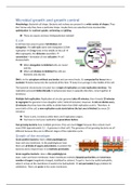
Microbial growth and growth control
Morphology: Bacterial cell shape. Bacteria and archaea are present in a wide variety of shapes. They
don’t know why they have a particular shape, maybe there are selective forces involved like
optimization for nutrient uptake, swimming and gliding.
There are square archaea (Haloquadratum walsbyi)
E.coli:
E.coli have two ways to grow: Cell division and
elongation. The cell cycle starts with elongation & DNA
replication Z ring forms in the middle of the cell
DNA segregates, the divisome assembles
constriction = formation of new cell poles cell
division/birth.
When elongation is inhibited cells are round
and die.
When cell division is inhibited the cells are
filaments and also die.
DNA is in the cytoplasm without any border and can move freely. It’s compacted by forces into a
nucleoid. Proteins bump into the nucleoid all the time keep it on average in the middle of the cell.
The bacterial chromosome (circular) has 1 origin of replication and one replication terminus. The
replication proceeds bidirectionally (2 polymerases leave in opposite direction, come together at
terminus).
Multiple fork replication: Replication of circular genome takes 40 minutes, then it needs 20 minutes
to segregate the genome in two daughter cells (=total 60 minutes). However, E.coli can divide every
20 minutes thus they have the ability to divide faster than DNA replication cycle is. Therefore, in
every birth of the cell, a new replication cycle starts before the last one is finished (so every 20
minutes).
There is only 1 terminus while there are 8 replication origins.
You have to start your replication 2 generations before.
Fast growing bacteria have multiple genomes. They are much bigger because they contain much
more DNA (approximately 3,5 – 4 genomes in their cell). The genomes of fast growing bacteria are all
different because they are in different stages of the replication cycle.
Growth of the envelope:
Gram positive bacteria: Have a thick peptidoglycan
layer and one membrane. In the peptidoglycan layer
there are all kinds of sugars and proteins, to protect the
bacteria and to import food. It is relatively permeable.
Gram negative bacteria: Have a thin peptidoglycan
layer, outer and inner membrane. Outer membrane contains lipopolysaccharides and enormous
number of sugars (negatively charged, stabilized by cations). To get in, has to be really hydrophilic,
when it arrives at the membrane it needs to be hydrophobic not permeable (huge problem for
antibiotics). There are pores in the outer membrane.
,Gram staining: Dye binds to peptidoglycan, binds good to gram positive bacteria.
Peptidoglycan layer determines shape. Peptidoglycan is a closed network of sugar chains covalently
linked by peptide side chains. It also protects the bacteria against internal pressure. Peptidoglycan is
a crosslinked polymer of 2 sugars and a side chain peptide.
Sugars are linked to each other by transglycosylase reaction.
Sugars are linked to the peptide by transpeptidase reaction.
Penicillin binding proteins (PBPs): PBPs are the enzyme that
make peptidoglycan by coupling sugars to each other
(transglycosylation) and by crosslinking peptide side chains
to each other (transpeptidation). They bind to the acceptor
and donor stempeptide. The donor stempeptide has the D-
ala-D-ala sequence. The last D-ala is cleaved off to provide
energy for covalent linkage between Dap and D-ala.
Penicillin is similar to D-ala-D-ala and binds to active site serine of penicillin binding proteins
covalent bond molecule cannot function anymore. Penicillin inhibits PBPs since the
molecule is stuck.
Penicillin kill all growing bacteria since it inhibits transpeptidation only sugars can be
added to each other weak. Infection can suddenly come back non growing bacteria can
persist and suddenly start growing again.
Ampicillin also inhibits this reaction.
Composition peptidoglycan: Peptidoglycan consists of two sugars of which one has small peptide
sidechains of 5 amino acids. These include D-amino acids and Dap (diaminopimeline acid, unique
amino acid in bacteria).
Important to recognise the D amino acids because our cells (normal proteases) can’t degrade
it.
Synthesis peptidoglycan: Precursor of peptidoglycan (lipid II) is synthesised in cytoplasm. It is then
coupled to a lipid tail and flipped across the membrane by MurJ. On the outside, new peptidoglycan
units are added by glycosyltransferase. Release of phosphate (donor, D-ala) is used as energy to
make the bond.
Two modes of peptidoglycan synthesis:
Elongation of length growth: By random insertion of material in the cylindrical part of the
cell. MreB (associated with MreC and MreD) localises as interrupted helix in E.coli (without
MreB they are spherical). Structure is similar to actin, both bind ATP and use it to
polymerase and depolymerise. PBPs insert new material in the peptidoglycan layer and push
the MreB helix backwards. The helix makes sure that this happens neatly spread over the
surface of the cell.
Cell division: Complete new synthesis of the new cell poles. When all the DNA is replicated
FtsZ polymerises in a ring like structure in the middle of the cell. FtsZ structure is similar to
tubulin, binding of GTP causes polymerisation. There is continuous assembly and
disassembly.
, How is the mid cell position determined? There is always enough FtsZ to polarise, E.coli has chosen
to inhibit FtsZ polymerisation instead of the amount of FtsZ. Why does it make only one ring in the
middle of the cell at the time the cell divides? Multiple reasons:
1. Presence of the nucleoid: The SlmA (NOC) protein is bound to the DNA, this interferes with
FtsZ polymerisation. Therefore, FtsZ will not easily polymerize in the vicinity of an active
transcribing nucleoid.
2. MinCDE system: Prevents polymerization in the vicinity of the cell poles. MinCD(ATP) binds
to the cell pole and inhibits FtsZ polymerization at that pole. MinE stimulates the ATPase of
MinD (so ATP ADP). MinCD(ADP) does not bind to the cell pole so you get release from
the membrane into the cytoplasm gradient, MinD and MinC move to the opposite pole.
On the way, cytosolic MinCD binds ATP again, causing it to bind to the opposite pole. On
average, FtsZ cannot polymerize into a Z-ring at the poles. You get MinCD oscillation.
Min system oscillation is detected by fusing proteins to GFP. Absence of Min system and local
minimum in DNA (NOC) allows FtsZ polymerisation at the middle.
FtsZ is not the only protein involved in cell division. FtsZ recruits other proteins to help. These are
responsible for the synthesis of the new cell poles during cell division.
Time scale to assembly of the complex/divisome:
Exponential phase: Typically the healthiest cells.
Stationary phase: Growth rate of population is zero.
Either the essential nutrient is used up or waste product
of the organism accumulates in the medium.
Steady state growth: Metabolism of the cells is constant and
they experience the environment as constant. Constant average
mass = constant age frequency distribution. Therefore cell length is cell age
(straight line).
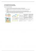
Immunolabeling: To find out at what timepoint these proteins localise in the cell. Fix
cell permeabilise add fluorescent labelled antibodies that recognise the ring
look under microscope image analysis (accumulation FtsZ and cell length).
Correlate cell length to cell age. Ring forms at 40% of cell age.
Left: Cell length.
Right: Amount of fluorescence in middle cell against rest of fluorescence.
MreB recruits the protein complex/elongasome to synthesise the envelope during
lateral growth. After about 30-40% of the cell cycle, the FtsZ ring with some
associated proteins form at mid cell. At 60% cell cycle all other proteins have
assembled into the divisome start division & synthesis of new cell poles directed
by treadmilling of FtsZ.
FtsZ is treadmilling: Binding GTP (polymerizes) on 1 side, losing molecules (depolymerizes)
on the other side. While moving in circles it drags the peptidoglycan synthesizing protein
complexes along (or other way around?). Speed is dependent on GTP hydrolysis.
Candidatus Thiosymbion oneisti: Symbiont with the nematode Laxus oneistus, the worm is covered
in it (bacterial coat). All bacteria have the same length, they grow in with and divide in cell length
Morphology: Bacterial cell shape. Bacteria and archaea are present in a wide variety of shapes. They
don’t know why they have a particular shape, maybe there are selective forces involved like
optimization for nutrient uptake, swimming and gliding.
There are square archaea (Haloquadratum walsbyi)
E.coli:
E.coli have two ways to grow: Cell division and
elongation. The cell cycle starts with elongation & DNA
replication Z ring forms in the middle of the cell
DNA segregates, the divisome assembles
constriction = formation of new cell poles cell
division/birth.
When elongation is inhibited cells are round
and die.
When cell division is inhibited the cells are
filaments and also die.
DNA is in the cytoplasm without any border and can move freely. It’s compacted by forces into a
nucleoid. Proteins bump into the nucleoid all the time keep it on average in the middle of the cell.
The bacterial chromosome (circular) has 1 origin of replication and one replication terminus. The
replication proceeds bidirectionally (2 polymerases leave in opposite direction, come together at
terminus).
Multiple fork replication: Replication of circular genome takes 40 minutes, then it needs 20 minutes
to segregate the genome in two daughter cells (=total 60 minutes). However, E.coli can divide every
20 minutes thus they have the ability to divide faster than DNA replication cycle is. Therefore, in
every birth of the cell, a new replication cycle starts before the last one is finished (so every 20
minutes).
There is only 1 terminus while there are 8 replication origins.
You have to start your replication 2 generations before.
Fast growing bacteria have multiple genomes. They are much bigger because they contain much
more DNA (approximately 3,5 – 4 genomes in their cell). The genomes of fast growing bacteria are all
different because they are in different stages of the replication cycle.
Growth of the envelope:
Gram positive bacteria: Have a thick peptidoglycan
layer and one membrane. In the peptidoglycan layer
there are all kinds of sugars and proteins, to protect the
bacteria and to import food. It is relatively permeable.
Gram negative bacteria: Have a thin peptidoglycan
layer, outer and inner membrane. Outer membrane contains lipopolysaccharides and enormous
number of sugars (negatively charged, stabilized by cations). To get in, has to be really hydrophilic,
when it arrives at the membrane it needs to be hydrophobic not permeable (huge problem for
antibiotics). There are pores in the outer membrane.
,Gram staining: Dye binds to peptidoglycan, binds good to gram positive bacteria.
Peptidoglycan layer determines shape. Peptidoglycan is a closed network of sugar chains covalently
linked by peptide side chains. It also protects the bacteria against internal pressure. Peptidoglycan is
a crosslinked polymer of 2 sugars and a side chain peptide.
Sugars are linked to each other by transglycosylase reaction.
Sugars are linked to the peptide by transpeptidase reaction.
Penicillin binding proteins (PBPs): PBPs are the enzyme that
make peptidoglycan by coupling sugars to each other
(transglycosylation) and by crosslinking peptide side chains
to each other (transpeptidation). They bind to the acceptor
and donor stempeptide. The donor stempeptide has the D-
ala-D-ala sequence. The last D-ala is cleaved off to provide
energy for covalent linkage between Dap and D-ala.
Penicillin is similar to D-ala-D-ala and binds to active site serine of penicillin binding proteins
covalent bond molecule cannot function anymore. Penicillin inhibits PBPs since the
molecule is stuck.
Penicillin kill all growing bacteria since it inhibits transpeptidation only sugars can be
added to each other weak. Infection can suddenly come back non growing bacteria can
persist and suddenly start growing again.
Ampicillin also inhibits this reaction.
Composition peptidoglycan: Peptidoglycan consists of two sugars of which one has small peptide
sidechains of 5 amino acids. These include D-amino acids and Dap (diaminopimeline acid, unique
amino acid in bacteria).
Important to recognise the D amino acids because our cells (normal proteases) can’t degrade
it.
Synthesis peptidoglycan: Precursor of peptidoglycan (lipid II) is synthesised in cytoplasm. It is then
coupled to a lipid tail and flipped across the membrane by MurJ. On the outside, new peptidoglycan
units are added by glycosyltransferase. Release of phosphate (donor, D-ala) is used as energy to
make the bond.
Two modes of peptidoglycan synthesis:
Elongation of length growth: By random insertion of material in the cylindrical part of the
cell. MreB (associated with MreC and MreD) localises as interrupted helix in E.coli (without
MreB they are spherical). Structure is similar to actin, both bind ATP and use it to
polymerase and depolymerise. PBPs insert new material in the peptidoglycan layer and push
the MreB helix backwards. The helix makes sure that this happens neatly spread over the
surface of the cell.
Cell division: Complete new synthesis of the new cell poles. When all the DNA is replicated
FtsZ polymerises in a ring like structure in the middle of the cell. FtsZ structure is similar to
tubulin, binding of GTP causes polymerisation. There is continuous assembly and
disassembly.
, How is the mid cell position determined? There is always enough FtsZ to polarise, E.coli has chosen
to inhibit FtsZ polymerisation instead of the amount of FtsZ. Why does it make only one ring in the
middle of the cell at the time the cell divides? Multiple reasons:
1. Presence of the nucleoid: The SlmA (NOC) protein is bound to the DNA, this interferes with
FtsZ polymerisation. Therefore, FtsZ will not easily polymerize in the vicinity of an active
transcribing nucleoid.
2. MinCDE system: Prevents polymerization in the vicinity of the cell poles. MinCD(ATP) binds
to the cell pole and inhibits FtsZ polymerization at that pole. MinE stimulates the ATPase of
MinD (so ATP ADP). MinCD(ADP) does not bind to the cell pole so you get release from
the membrane into the cytoplasm gradient, MinD and MinC move to the opposite pole.
On the way, cytosolic MinCD binds ATP again, causing it to bind to the opposite pole. On
average, FtsZ cannot polymerize into a Z-ring at the poles. You get MinCD oscillation.
Min system oscillation is detected by fusing proteins to GFP. Absence of Min system and local
minimum in DNA (NOC) allows FtsZ polymerisation at the middle.
FtsZ is not the only protein involved in cell division. FtsZ recruits other proteins to help. These are
responsible for the synthesis of the new cell poles during cell division.
Time scale to assembly of the complex/divisome:
Exponential phase: Typically the healthiest cells.
Stationary phase: Growth rate of population is zero.
Either the essential nutrient is used up or waste product
of the organism accumulates in the medium.
Steady state growth: Metabolism of the cells is constant and
they experience the environment as constant. Constant average
mass = constant age frequency distribution. Therefore cell length is cell age
(straight line).
Immunolabeling: To find out at what timepoint these proteins localise in the cell. Fix
cell permeabilise add fluorescent labelled antibodies that recognise the ring
look under microscope image analysis (accumulation FtsZ and cell length).
Correlate cell length to cell age. Ring forms at 40% of cell age.
Left: Cell length.
Right: Amount of fluorescence in middle cell against rest of fluorescence.
MreB recruits the protein complex/elongasome to synthesise the envelope during
lateral growth. After about 30-40% of the cell cycle, the FtsZ ring with some
associated proteins form at mid cell. At 60% cell cycle all other proteins have
assembled into the divisome start division & synthesis of new cell poles directed
by treadmilling of FtsZ.
FtsZ is treadmilling: Binding GTP (polymerizes) on 1 side, losing molecules (depolymerizes)
on the other side. While moving in circles it drags the peptidoglycan synthesizing protein
complexes along (or other way around?). Speed is dependent on GTP hydrolysis.
Candidatus Thiosymbion oneisti: Symbiont with the nematode Laxus oneistus, the worm is covered
in it (bacterial coat). All bacteria have the same length, they grow in with and divide in cell length