1 - FUNCTIONAL NEUROANATOMY OF VOLUNTARY
MOTOR CONTROL
Locus à a small, well-defined group of cells
Ganglion à a collection of neurons in the PNS
Substantia à a group of related neurons deep within the brain, usually with less distinct borders than
those of nuclei
Nucleus à a clearly defined mass of neurons, usually deep within the brain
Tract à collection of CNS axons having a common site of origin and a common destination
Bundle à a collection of axons that run together but do not necessarily have the same origin and
destination
Capsule à a collection of axons that connect the cerebrum with the brain stem
BEAR - THE STRUCTURE OF THE NERVOUS SYSTEM
THE CENTRAL NERVOUS SYSTEM
CNS consists of parts of the NS in the brain and spinal cord (both encased in bone)
Cerebrum • Rostral-most and largest part of the brain
• Clearly split down the middle into 2 cerebral hemispheres. Separated by the deep sagittal
fissure
• Right cerebral hemisphere receives sensations from, and controls movements of, the left side
of body
Cerebellum • Contains as many neurons as both cerebral hemispheres combined
• Left side of cerebellum concerned with movements on left side of body (Ipsilateral)
Brain stem • Forms the stalk from which the cerebral hemispheres and cerebellum sprout
• Serves to relay information from cerebrum to spinal cord and cerebellum & vice versa
Spinal cord • Is encased in the bony vertebral column and is attached to the brain stem
• A transection of the spinal cord results in anesthesia in the skin and paralysis of the muscles
caudal to the cut
• Communicates via spinal nerves (PNS) that exit the spinal cord through notches between
each vertebra of the vertebral column
• Each spinal nerve attaches to spinal cord via 2 branches: dorsal root and ventral root
• Dorsal root – contains axons bringing information INTO the spinal cord
• Ventral root – contains axons carrying information AWAY from the spinal cord
,THE PERIPHERAL NERVOUS SYSTEM
1. Somatic PNS, 2. Visceral PNS
Somatic PNS Visceral PNS (= autonomous PNS)
• All spinal nerves that innervate the skin, the • Consists of neurons that innervate the
joints, and the muscles that are under internal organs, blood vessels, glands
voluntary control are part of the somatic • Visceral sensory axons bring information
PNS about visceral function to the CNS
• Somatic motor axons command muscle • Ex. Visceral motor fibers command the
contraction, derive from motor neurons in contraction / relaxation of muscles that
ventral spinal cord form walls of intestines
• Somatic sensory axons enter the spinal
cord via the dorsal roots
• Cell bodies of these neurons lie outside the
spinal cord in clusters à dorsal root
ganglia
Afferent neurons à carry to
Efferent neurons à carry from
THE CRANIAL NERVES
• There’s 12 pairs of cranial nerves that arise from the brain stem and innervate the head
• Many cranial nerves contain a complex mixture of axons that perform different functions
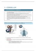
THE MENINGES
• The CNS does not come into direct contact with the overlying bone - protected by
the meninges
• Outermost à dura mater
• Mid à arachnoid membrane (spider web-like)
• Bottom à pia mater (blood vessels)
UNDERSTANDING CNS STRUCTURE THROUGH DEVELOPMENT
The entire CNS is derived from the walls of neural tube, which itself becomes the adult ventricular system
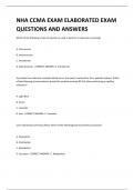
FORMATION OF THE NEURAL TUBE
• Embryo begins as a flat disk with 3 distinct layers of cells:
endoderm, mesoderm, ectoderm
1. Endoderm – lining of many internal organs
2. Mesoderm – bones of skeleton and muscles
3. Ectoderm – entire nervous system (neural plate) and
skin
, • Next: formation of neural groove that runs rostral to caudal. Walls of the groove are the neural
folds, which fuse dorsally and form the neural tube
o The entire nervous system develops from the walls of the neural tube
• As the neural folds come together, some neural ectoderm is pinched off and becomes the neural
crest
o All neurons with cell bodies in the PNS derive from the neural crest
• Neural crest develops in close association with the underlying mesoderm
• Mesoderm forms bulges on either side of the neural tube – somites. From these somites, the 33
individual vertebrae of the spinal column and skeletal muscles develop
• Neurulation – process by which the neural plate becomes the neural tube (very early in embryonic
development, 22 days after conception)
Nutrition and the Neural Tube
• Neural tube formation is essential for early nervous system development, occurring about 3
weeks after conception.
• Neural tube defects (NTDs) occur if the tube doesn’t close properly, affecting about 1 in 500 live
births.
• Many NTDs are linked to low folic acid (vitamin
B9) intake in the mother's diet shortly after
conception. Folic acid supplementation could
reduce NTDs by up to 90%.
• Failure of tube closure in the anterior region
causes anencephaly (fatal due to brain and skull
degeneration).
• Failure in the posterior region causes spina bifida
(non-fatal but may need extensive medical care).
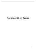
THREE PRIMARY BRAIN VESICLES
• Differentiation – process by which structures become more complex and functionally specialized
during development
• First step in differentiation à development of the 3 primary vesicles (at rostral end of neural tube)
o The entire brain derives from the three primary vesicles of the neural tube
• Prosencephalon (forebrain) - rostral-most vesicle
• Mesencephalon (midbrain) – behind prosencephalon
• Rhombencephalon (hindbrain) – caudal to mesencephalon, connects with caudal neural tube,
giving rise to spinal cord
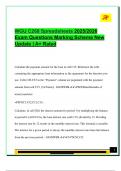
PUTTING THE PIECES TOGETHER
• The paired hemispheres of the telencephalon
surround the lateral ventricles
• Ventral and lateral to the lateral ventricles lies the
basal telencephalon
• The lateral ventricles are continuous with the third
ventricle of the diencephalon
MOTOR CONTROL
Locus à a small, well-defined group of cells
Ganglion à a collection of neurons in the PNS
Substantia à a group of related neurons deep within the brain, usually with less distinct borders than
those of nuclei
Nucleus à a clearly defined mass of neurons, usually deep within the brain
Tract à collection of CNS axons having a common site of origin and a common destination
Bundle à a collection of axons that run together but do not necessarily have the same origin and
destination
Capsule à a collection of axons that connect the cerebrum with the brain stem
BEAR - THE STRUCTURE OF THE NERVOUS SYSTEM
THE CENTRAL NERVOUS SYSTEM
CNS consists of parts of the NS in the brain and spinal cord (both encased in bone)
Cerebrum • Rostral-most and largest part of the brain
• Clearly split down the middle into 2 cerebral hemispheres. Separated by the deep sagittal
fissure
• Right cerebral hemisphere receives sensations from, and controls movements of, the left side
of body
Cerebellum • Contains as many neurons as both cerebral hemispheres combined
• Left side of cerebellum concerned with movements on left side of body (Ipsilateral)
Brain stem • Forms the stalk from which the cerebral hemispheres and cerebellum sprout
• Serves to relay information from cerebrum to spinal cord and cerebellum & vice versa
Spinal cord • Is encased in the bony vertebral column and is attached to the brain stem
• A transection of the spinal cord results in anesthesia in the skin and paralysis of the muscles
caudal to the cut
• Communicates via spinal nerves (PNS) that exit the spinal cord through notches between
each vertebra of the vertebral column
• Each spinal nerve attaches to spinal cord via 2 branches: dorsal root and ventral root
• Dorsal root – contains axons bringing information INTO the spinal cord
• Ventral root – contains axons carrying information AWAY from the spinal cord
,THE PERIPHERAL NERVOUS SYSTEM
1. Somatic PNS, 2. Visceral PNS
Somatic PNS Visceral PNS (= autonomous PNS)
• All spinal nerves that innervate the skin, the • Consists of neurons that innervate the
joints, and the muscles that are under internal organs, blood vessels, glands
voluntary control are part of the somatic • Visceral sensory axons bring information
PNS about visceral function to the CNS
• Somatic motor axons command muscle • Ex. Visceral motor fibers command the
contraction, derive from motor neurons in contraction / relaxation of muscles that
ventral spinal cord form walls of intestines
• Somatic sensory axons enter the spinal
cord via the dorsal roots
• Cell bodies of these neurons lie outside the
spinal cord in clusters à dorsal root
ganglia
Afferent neurons à carry to
Efferent neurons à carry from
THE CRANIAL NERVES
• There’s 12 pairs of cranial nerves that arise from the brain stem and innervate the head
• Many cranial nerves contain a complex mixture of axons that perform different functions
THE MENINGES
• The CNS does not come into direct contact with the overlying bone - protected by
the meninges
• Outermost à dura mater
• Mid à arachnoid membrane (spider web-like)
• Bottom à pia mater (blood vessels)
UNDERSTANDING CNS STRUCTURE THROUGH DEVELOPMENT
The entire CNS is derived from the walls of neural tube, which itself becomes the adult ventricular system
FORMATION OF THE NEURAL TUBE
• Embryo begins as a flat disk with 3 distinct layers of cells:
endoderm, mesoderm, ectoderm
1. Endoderm – lining of many internal organs
2. Mesoderm – bones of skeleton and muscles
3. Ectoderm – entire nervous system (neural plate) and
skin
, • Next: formation of neural groove that runs rostral to caudal. Walls of the groove are the neural
folds, which fuse dorsally and form the neural tube
o The entire nervous system develops from the walls of the neural tube
• As the neural folds come together, some neural ectoderm is pinched off and becomes the neural
crest
o All neurons with cell bodies in the PNS derive from the neural crest
• Neural crest develops in close association with the underlying mesoderm
• Mesoderm forms bulges on either side of the neural tube – somites. From these somites, the 33
individual vertebrae of the spinal column and skeletal muscles develop
• Neurulation – process by which the neural plate becomes the neural tube (very early in embryonic
development, 22 days after conception)
Nutrition and the Neural Tube
• Neural tube formation is essential for early nervous system development, occurring about 3
weeks after conception.
• Neural tube defects (NTDs) occur if the tube doesn’t close properly, affecting about 1 in 500 live
births.
• Many NTDs are linked to low folic acid (vitamin
B9) intake in the mother's diet shortly after
conception. Folic acid supplementation could
reduce NTDs by up to 90%.
• Failure of tube closure in the anterior region
causes anencephaly (fatal due to brain and skull
degeneration).
• Failure in the posterior region causes spina bifida
(non-fatal but may need extensive medical care).
THREE PRIMARY BRAIN VESICLES
• Differentiation – process by which structures become more complex and functionally specialized
during development
• First step in differentiation à development of the 3 primary vesicles (at rostral end of neural tube)
o The entire brain derives from the three primary vesicles of the neural tube
• Prosencephalon (forebrain) - rostral-most vesicle
• Mesencephalon (midbrain) – behind prosencephalon
• Rhombencephalon (hindbrain) – caudal to mesencephalon, connects with caudal neural tube,
giving rise to spinal cord
PUTTING THE PIECES TOGETHER
• The paired hemispheres of the telencephalon
surround the lateral ventricles
• Ventral and lateral to the lateral ventricles lies the
basal telencephalon
• The lateral ventricles are continuous with the third
ventricle of the diencephalon