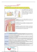
Test bank clinical manifestation and
assessment of respiratory disease
, Table of Contents
PART I: ASSESSMENT OF CARDIOPULMONARY DISEASE
SECTION I: Bedside Diagnosis
1.The Patient Interview
2.The Physical Examination
3.The Pathophysiologic Basis for Common Clinical Manifestations
SECTION II: Clinical Data Obtained from Laboratory Tests and Special
Procedures — Objective Findings
4.Pulmonary Function Testing
5.Blood Gas Assessment
6.Assessment of Oxygenation
7.Assessment of the Cardiovascular System
8.Radiologic Examination of the Chest
9.Other Important Tests and Procedures
SECTION III: The Therapist-Driven Protocol Program — The Essentials
10.The Therapist-Driven Protocol Program
11.Respiratory Failure and Ventilatory Management Protocols
12.Recording Skills and Intra-Professional Communication
,PART II: OBSTRUCTIVE LUNG DISEASE
13.Chronic Obstructive Pulmonary Disease, Chronic Bronchitis, and Emphysema
14.Asthma
15.Cystic Fibrosis
16.Bronchiectasis
PART III: LOSS OF ALVEOLAR VOLUME
17.Atelectasis
PART IV: INFECTIOUS PULMONARY DISEASE
18.Pneumonia, Lung Abscess Formation, and Important Fungal Diseases
19.Tuberculosis
PART V: PULMONARY VASCULAR DISEASE
20.Pulmonary Edema
21.Pulmonary Vascular Disease: Pulmonary Embolism and Pulmonary Hypertension
PART VI: CHEST AND PLEURAL TRAUMA
22.Flail Chest
23.Pneumothorax
PART VII: DISORDERS OF THE PLEURA AND THE CHEST WALL
24.Pleural Effusion and Empyema
25.Kyphoscoliosis
, PART VIII: LUNG CANCER
26.Cancer of the Lung
PART IX: ENVIRONMENTAL LUNG DISEASES
27.Interstitial Lung Diseases
PART X: DIFFUSE ALVEOLAR DISEASE
28.Acute Respiratory Distress Syndrome
PART XI: NEURO-RESPIRATORY DISORDERS
29.Guillain-Barre Syndrome
30.Myasthenia Gravis
31.Cardiopulmonary Assessment and Care of Patients with Neuromuscular Disease
PART XII: SLEEP-RELATED BREATHING DISORDERS
32.Sleep Apnea
PART XIII: NEWBORN AND EARLY CHILDHOOD
CARDIOPULMONARY DISORDERS
33.Newborn Assessment and Management
34.Pediatric Assessment and Management
35.Meconium Aspiration Syndrome
36.TrANS ient Tachypnea of the Newborn
37.Respiratory Distress Syndrome