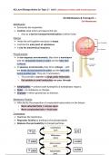
AS Level Biology Notes for Topic 3 – Unit 2 ; Development, Biodiversity & Conservation
3A Cell Structure —-
Observing Cells
➢ Magnification → a measure of how much bigger the image you see is than the
real object.
➢ A light microscope has 2 types of lenses; an eyepiece lens of x10 and a series
of 3 objective lenses with different magnifications each.
○ Total magnification = Eyepiece lens magnification x Objective lens
magnification
➢ Resolution (resolving power) → a measure of how close 2 objects should be
before they’re seen as one. This is the ability to distinguish between two different
points.
➢ The resolution of a microscope limits the magnification that it’s capable of.
○ The resolution of a light microscope is limited by the wavelength of light.
○ Electron microscopes have a higher resolution as electrons have a much
smaller wavelength than visible light.
Light Microscopes: used for specimens larger than 200nm
➢ They shine light through the specimen – they’re useful for looking at whole cells,
small organisms and tissues within organs.
○ They’re cheap & portable, easy to use, can view whole & living specimens.
○ However, it only shows 2D images, requires staining which can introduce
artefacts, has a resolving power of 200nm and a magnification of x1500.
Electron Microscopes: used for specimens larger than 0.5nm
➢ The fired electrons are picked up by an electromagnetic lens to produce an image
– it requires the specimen to be dead, providing a snapshot. There are 2 types:
➢ Transmission Electron Microscopes (TEM) → transmit a beam through a thin
specimen and used to produce 2D images of smaller sub-cellular ultrastructures.
○ Can see ultrastructures in detail, has a resolving power of 0.1nm and a
magnification of x2,000,000
➢ Scanning Electron Microscopes (SEM) → scans a fire beam of electrons and
collects the electrons scattered on the surface, used to produce 3D images of
the specimens.
○ Has a resolving power of 20nm and a magnification of x200,000.
○ However, specimens must be fixed in plastic and viewed in a vacuum (dead),
specimens may get damaged by the beam, specimens must be stained with
heavy metals which introduced artefacts, they are expensive and large.
, AS Level Biology Notes for Topic 3 – Unit 2 ; Development, Biodiversity & Conservation
➢ Sometimes, specimens must be stained as the cytoplasm/cell structures are
transparent or difficult to distinguish.
○ Haematoxylin — stains plant/animal cells’ nuclei purple/blue.
○ Methylene blue — stains animal cells' nuclei blue.
○ Acetocarmine — stains chromosomes in dividing nuclei of cells.
○ Iodine — stains starch-containing material in plant cells blue-black.
○ Toluidine blue — stains tissues that contain DNA/RNA blue.
Cell Theory:
➢ All living organisms are made up of one or more cells.
➢ Cells are the basic functional unit in living organisms.
➢ New cells are produced from pre-existing cells.
➢ All cells share common features; cell surface membranes, cytoplasm, DNA and
ribosomes.
➢ Ultrastructure → internal structures of the cell.
Eukaryotic Cells – Common Cellular Structures
All cells are surrounded by a membrane which controls the
Cell Surface
exchange of materials between the internal and external
Membrane
environment.
Relatively large and separated from the cytoplasm by a
double membrane (nuclear envelope) which has many
pores that allow mRNA and enzymes to enter/exit.
Nucleus The nucleus also has chromatin (material for chromosomes),
chromosomes are sections of linear DNA tightly wound
around histones – darkly stained regions of the nucleus are
the nucleolus, which are sites of ribosome production.
The site of aerobic respiration, they’re surrounded by a
double membrane with the inner membrane folded into
cristae. The matrix contains enzymes needed for aerobic
Mitochondria
respiration, producing ATP; mitochondrial DNA and
ribosomes are also found in the matrix (needed for the
replication of mitochondria before cell division).
Found on the RER/cytoplasm and are not surrounded by a
membrane, they’re made up of ribosomal RNA and
Ribosomes proteins. In prokaryotes, they’re 70s (50s large/30s small) &
in eukaryotes, they’re 80s (60s large/40s small)
They’re the site of protein synthesis (translation).