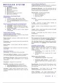
Actin and Myosin Myofilaments
MUSCULAR SYSTEM Troponin molecules – binding sites for Ca2+; attached at
Functions (M2RPC3) specific intervals along the actin myofilaments
1. Movement of the body.
2. Maintenance of posture. Tropomyosin filaments – cover the attachment sites on
3. Respiration the actin myofilaments; located along the grove bet. the
4. Production of body heat twisted strands of actin myofilaments
5. Communication
6. Constriction of organs and vessels Myosin heads – resemble golf club heads;
7. Contraction of the heart Ø Bind to attachment sites
Ø Bend and straighten
Characteristics of the Skeletal Muscle Ø Break down ATP
Skeletal Muscle
§ Constitutes approx. 40% of body weight Sarcomere
§ Muscles are attached to the skeletal system § Basic structural and functional unit of skeletal
§ Also called Striated Muscle; transverse bands or muscle
striations
Z disk – network of protein fibers forming an
Major Functional Characteristics of Skeletal Muscle attachment site for actin myofilaments
1. Contractility – ability to shorten with force
2. Excitability – capacity to respond to a stimulus I band – consists of actin myofilaments; spans each Z
3. Extensibility – ability to be stretched to their disk
normal resting length
4. Elasticity – ability to recoil to their original A band – darker, central region that extends the length
resting length of the myosin of myofilaments
Skeletal Muscle Structure H zone – second light zone that consists of myosin
Connective Tissue Coverings of Muscle myofilaments
Epimysium/Muscular fascia – connective tissue sheath
that surrounds a skeletal muscle M line – dark-staining bands
Muscle fasciculi – numerous visible bundles that make The arrangement of the actin and myosin filaments in
up the muscle sacromeres gives the myofibrils a banded appearance.
Perimysium – loose connective tissue that surrounds the The alternating I bands and A bands of the sacromeres
muscle fasciculi are responsible for the striations in the skeletal muscle
fibers.
Muscle fibers – several muscle cells that composes a
fasciculus Excitability of Muscle Fibers
Resting membrane potential – cell membranes have a
Endomysium – loose connective tissue that surrounds a negative charge on the inside relative to a positive
muscle fiber charge outside; occurs bcos there is an uneven
distribution of ions
Muscle Fiber Structure 1. Concentration of K+ inside the cell CM >
Sarcolemma – cell membrane of the muscle fiber outside the CM
2. Concentration of Na+ outside the CM > inside
Transverse tubules (T tubules) – tube-like invaginations the CM
w/c occur at regular intervals along the muscle fiber
Different types of Ion Channels
Sarcoplasmic reticulum – highly organized smooth E.R.; Ø Nongated/Leak channels – always open
has a relatively high concentration of Ca2+ (muscle
contraction) Ø Chemically gated channels – closed until a
chemical binds them and stimulates them to
Sarcoplasm – cytoplasm of a muscle fiber open
Myofibrils – threadlike structures composed of: Depolarization – the inside of the CM membrane comes
Ø Actin Myofilaments (thin filaments; purple) more positive than the outside of the cell; Na+ ions
Ø Myosin Myofilaments (thick filaments; green) move into cells
Sarcomere – highly ordered, repeating units of actin + Repolarization – the change back to the resting
myosin myofilaments; joined end to end to form the membrane potential; K+ ions moves out of cells
myofibril
Action Potentials – the rapid depolarization and
repolarization of the CM; results in muscle contraction
M o r a n o , M . A .
, Nerve Supply Aerobic Respiration – requires O2; breaks down glucose
Motor neurons – specialized nerve cells that stimulate to produce ATP, CO2, H2O
muscles to contract
Anaerobic respiration – doesn’t require O2; breaks
Neuromuscular junction – a branch that forms a down glucose to yield ATP and lactic acid
junction with a muscle fiber
Creatine phosphate – high-energy molecule that can be
Synapse – cell-to-cell junction bet. a nerve cell and stored in muscle fibers
another nerve cell/effector cell
Fatigue
Motor unit – a single motor neuron and all the skeletal § A state of reduced work capacity
muscle fibers it innervates
Muscular Fatigue – when muscle fibers use ATP faster
Presynaptic terminal – enlarged axon terminal than they are produced; when the effectiveness of Ca+ to
stimulate actin + myosin is reduced
Synaptic cleft – the space bet. the presynaptic terminal
and the muscle fiber membrane Physiological contracture – muscles may become
incapable of either contracting or relaxing
Postsynaptic membrane – the muscle fiber membrane
Psychological fatigue – involves the CNS; an individual
Synaptic vesicles – presynaptic terminal that contains perceives that continued muscle contraction is
small vesicles impossible
Acetylcholine (ACh) – neurotransmitter contained in Type of Muscle Contractions
the vesicles; a molecule released by a presynaptic nerve Isometric contractions – equal distance; length of the
cell that stimulates/inhibits a postsynaptic cell muscle does not change; the amount of tension increases
during the contraction process
Acetylcholinesterase – an enzyme that rapidly breaks
down the synaptic cleft bet. the neuron and the muscle Isotonic contraction – equal tension; the amount of
fiber tension produced by the muscle is constant during
contraction; length of the muscle decreases
Muscle Contraction Ø Cocentric contractions – isotonic; muscle
Sliding filament model – sliding of actin myofilaments tension increases as the muscle shortens
past myosin myofilaments during contraction Ø Eccentric contractions – isotonic; tension is
maintained in a muscle; the opposing resistance
Cross-bridges – myosin heads attach to the myosin causes the muscle to lengthen
attachment sites on the actin myofilaments
Muscle Tone
Muscle Twitch, Summation, Tetanus, Recruitment § Constant tension produced by body muscles
Muscle Twitch – contraction of a muscle fiber in over long periods of time
reponse to a stimulus § Responsible for keeping the back and legs
1. Lag/Latent Phase – time bet. the application of a straight, the head in an upright position, and the
stimulus and the beginning of contraction abdomen from bulging
2. Contraction Phase – time during which the
muscle contract Slow-Twitch and Fast-Twitch Fibers
3. Relaxation Phase – time during which the Classification of Muscle Fiber
muscle relaxes 1. Slow Twitch – contains type I myosin; contracts
slowly and resistant to fatigue respiration
Summation – the force of contraction of an individual
muscle fiber is increased by rapidly stimulating them 2.Fast Twitch
a. Type IIa – intermediate speed; more
Tetanus – convulsive tension; a sustained contraction fatigue resistant than type IIb
that occurs when the frequency of stimulus is so rapid b. Type IIb – contract 10x faster than type
that no relaxation occurs I
Ø Caused by Ca+ build up in the myofibrils Myglobin – stores oxygen temporarily
Recruitment – the no. of muscle fibers contraction is Hypertrophy – enlarging of muscle fibers
increased by the increasing no. of motor units stimulated
+ muscle contracts with more force Satellite cells – undifferentiated cells just below the
endomysium
Stimulus frequency – no. of times a motor neuron is
stimulated per second Smooth and Cardiac Muscle
Autorhythmicity – resulting periodic spontaneous
Energy Requirement for Muscle Contraction contraction of smooth muscle
M o r a n o , M . A .